 Movie
Movie Controller

[English] 日本語
 Yorodumi
- PDB-3f3m: Six Crystal Structures of Two Phosphopantetheine Adenylyltransfer... -
+ Open data

- Basic information
| Entry | Database: PDB / ID: 3f3m | ||||||
|---|---|---|---|---|---|---|---|
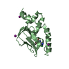
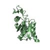
| Title | Six Crystal Structures of Two Phosphopantetheine Adenylyltransferases Reveal an Alternative Ligand Binding Mode and an Associated Structural Change | ||||||
 Components Components | Phosphopantetheine adenylyltransferase | ||||||
 Keywords Keywords | TRANSFERASE / Phosphopantetheine adenylyltransferase / PPAT / Coenzyme A biosynthetic pathway / Coenzyme A biosynthesis / Nucleotidyltransferase | ||||||
| Function / homology |  Function and homology information Function and homology informationpantetheine-phosphate adenylyltransferase / pantetheine-phosphate adenylyltransferase activity / coenzyme A biosynthetic process / ATP binding / cytoplasm Similarity search - Function | ||||||
| Biological species |   Staphylococcus aureus (bacteria) Staphylococcus aureus (bacteria) | ||||||
| Method |  X-RAY DIFFRACTION / SYNCHROTRON / MOLECULAR REPLACEMENT / Resolution: 2.4 Å X-RAY DIFFRACTION / SYNCHROTRON / MOLECULAR REPLACEMENT / Resolution: 2.4 Å | ||||||
 Authors Authors | Lee, H.H. / Yoon, H.J. / Suh, S.W. | ||||||
 Citation Citation | Journal: Acta Crystallogr.,Sect.F / Year: 2009 Title: The structure of Staphylococcus aureus phosphopantetheine adenylyltransferase in complex with 3'-phosphoadenosine 5'-phosphosulfate reveals a new ligand-binding mode Authors: Lee, H.H. / Yoon, H.J. / Kang, J.Y. / Park, J.H. / Kim, D.J. / Choi, K.H. / Lee, S.K. / Song, J. / Kim, H.J. / Suh, S.W. | ||||||
| History |
|
- Structure visualization
| Structure viewer | Molecule: MolmilJmol/JSmol |
|---|
- Downloads & links
-Download
| PDBx/mmCIF format | 3f3m.cif.gz | 46.6 KB | Display | PDBx/mmCIF format |
|---|---|---|---|---|
| PDB format | pdb3f3m.ent.gz | 32.1 KB | Display | PDB format |
| PDBx/mmJSON format | 3f3m.json.gz | Tree view | PDBx/mmJSON format | |
| Others |  Other downloads Other downloads |
-Validation report
| Arichive directory | https://data.pdbj.org/pub/pdb/validation_reports/f3/3f3mftp://data.pdbj.org/pub/pdb/validation_reports/f3/3f3m | HTTPS FTP |
|---|
-Related structure data
| Related structure data |  1b6tS  3f3i 3f3j 3f3l 3f3n 3f3o S: Starting model for refinement |
|---|---|
| Similar structure data |


-Links
 PDBj
PDBj
- Assembly
| Deposited unit | 
| ||||||||
|---|---|---|---|---|---|---|---|---|---|
| 1 |
| ||||||||
| Unit cell |
|
-Components
| #1: Protein | Mass: 19468.309 Da / Num. of mol.: 1 Source method: isolated from a genetically manipulated source Source: (gene. exp.) Staphylococcus aureus (bacteria) / Gene: coaD / Plasmid: pET-21a(+) / Production host: References: UniProt: P63820, pantetheine-phosphate adenylyltransferase |
|---|---|
| #2: Chemical | ChemComp-PPS /   Mass: 507.264 Da / Num. of mol.: 1 / Source method: obtained synthetically / Formula: C10H15N5O13P2S Mass: 507.264 Da / Num. of mol.: 1 / Source method: obtained synthetically / Formula: C10H15N5O13P2S |
| #3: Water | ChemComp-HOH /  Mass: 18.015 Da / Num. of mol.: 43 / Source method: isolated from a natural source / Formula: H2O Mass: 18.015 Da / Num. of mol.: 43 / Source method: isolated from a natural source / Formula: H2O |
-Experimental details
-Experiment
| Experiment | Method: X-RAY DIFFRACTION / Number of used crystals: 1 |
|---|
- Sample preparation
| Crystal | Density Matthews: 2.05 Å3/Da / Density % sol: 40.05 % |
|---|---|
| Crystal grow | Temperature: 297 K / Method: vapor diffusion, hanging drop / pH: 8 Details: 100mM imidazone, 0.9-1.2M potassium/sodium tartrate, 0.2M NaCl, pH8.0, VAPOR DIFFUSION, HANGING DROP, temperature 297K |
-Data collection
| Diffraction | Mean temperature: 100 K |
|---|---|
| Diffraction source | Source: SYNCHROTRON / Site: Photon Factory  / Beamline: BL-18B / Wavelength: 0.9788 Å / Beamline: BL-18B / Wavelength: 0.9788 Å |
| Detector | Type: ADSC QUANTUM 210 / Detector: CCD / Date: Feb 24, 2004 / Details: Mirrors |
| Radiation | Protocol: SINGLE WAVELENGTH / Monochromatic (M) / Laue (L): M / Scattering type: x-ray |
| Radiation wavelength | Wavelength: 0.9788 Å / Relative weight: 1 |
| Reflection | Resolution: 2.4→30 Å / Num. obs: 12227 / % possible obs: 98.8 % / Rmerge(I) obs: 0.112 / Net I/σ(I): 10.9 |
| Reflection shell | Resolution: 2.4→2.49 Å / Rmerge(I) obs: 0.397 / Mean I/σ(I) obs: 5.6 / Num. unique all: 1161 / % possible all: 93.9 |
- Processing
| Software |
| ||||||||||||||||||||||||||||
|---|---|---|---|---|---|---|---|---|---|---|---|---|---|---|---|---|---|---|---|---|---|---|---|---|---|---|---|---|---|
| Refinement | Method to determine structure: MOLECULAR REPLACEMENT Starting model: PDB ENTRY 1B6T Resolution: 2.4→30 Å / Occupancy max: 1 / Occupancy min: 1 / σ(F): 0
| ||||||||||||||||||||||||||||
| Solvent computation | Bsol: 34.131 Å2 | ||||||||||||||||||||||||||||
| Displacement parameters | Biso max: 95.89 Å2 / Biso mean: 31.96 Å2 / Biso min: 5.75 Å2
| ||||||||||||||||||||||||||||
| Refinement step | Cycle: LAST / Resolution: 2.4→30 Å
| ||||||||||||||||||||||||||||
| Refine LS restraints |
| ||||||||||||||||||||||||||||
| Xplor file |
|
