 Movie
Movie Controller

[English] 日本語
 Yorodumi
- PDB-1u7p: X-ray Crystal Structure of the Hypothetical Phosphotyrosine Phosp... -
+ Open data

- Basic information
| Entry | Database: PDB / ID: 1u7p | ||||||
|---|---|---|---|---|---|---|---|
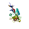
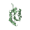
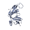
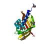
| Title | X-ray Crystal Structure of the Hypothetical Phosphotyrosine Phosphatase MDP-1 of the Haloacid Dehalogenase Superfamily | ||||||
 Components Components | magnesium-dependent phosphatase-1 | ||||||
 Keywords Keywords | HYDROLASE / HAD superfamily / phosphoryl transfer / phosphotyrosine phosphatase / aspartate nucleophile / enzyme evolution / structural enzymology / class III | ||||||
| Function / homology |  Function and homology information Function and homology informationfructosamine metabolic process / acid phosphatase activity / Hydrolases; Acting on ester bonds; Phosphoric-monoester hydrolases / phosphatase activity / protein-tyrosine-phosphatase / protein tyrosine phosphatase activity / metal ion binding Similarity search - Function | ||||||
| Biological species |  | ||||||
| Method |  X-RAY DIFFRACTION / MOLECULAR REPLACEMENT / Resolution: 1.9 Å X-RAY DIFFRACTION / MOLECULAR REPLACEMENT / Resolution: 1.9 Å | ||||||
 Authors Authors | Peisach, E. / Selengut, J.D. / Dunaway-Mariano, D. / Allen, K.N. | ||||||
 Citation Citation | Journal: Biochemistry / Year: 2004 Title: X-ray Crystal Structure of the Hypothetical Phosphotyrosine Phosphatase MDP-1 of the Haloacid Dehalogenase Superfamily Authors: Peisach, E. / Selengut, J.D. / Dunaway-Mariano, D. / Allen, K.N. #1: Journal: Biochemistry / Year: 2000Title: MDP-1: A novel eukaryotic magnesium-dependent phosphatase Authors: Selengut, J.D. / Levine, R.L. #2: Journal: Biochemistry / Year: 2001Title: MDP-1 is a new and distinct member of the haloacid dehalogenase family of aspartate-dependent phosphohydrolases Authors: Selengut, J.D. #3: Journal: Biochemistry / Year: 2000Title: The crystal structure of bacillus cereus phosphonoacetaldehyde hydrolase: insight into catalysis of phosphorus bond cleavage and catalytic diversification within the HAD enzyme superfamily Authors: Morais, M.C. / Zhang, W. / Baker, A.S. / Zhang, G. / Dunaway-Mariano, D. / Allen, K.N. | ||||||
| History |
|
- Structure visualization
| Structure viewer | Molecule: MolmilJmol/JSmol |
|---|
- Downloads & links
-Download
| PDBx/mmCIF format | 1u7p.cif.gz | 148 KB | Display | PDBx/mmCIF format |
|---|---|---|---|---|
| PDB format | pdb1u7p.ent.gz | 116.4 KB | Display | PDB format |
| PDBx/mmJSON format | 1u7p.json.gz | Tree view | PDBx/mmJSON format | |
| Others |  Other downloads Other downloads |
-Validation report
| Arichive directory | https://data.pdbj.org/pub/pdb/validation_reports/u7/1u7pftp://data.pdbj.org/pub/pdb/validation_reports/u7/1u7p | HTTPS FTP |
|---|
-Related structure data


-Links
 PDBj
PDBj
- Assembly
| Deposited unit | 
| ||||||||
|---|---|---|---|---|---|---|---|---|---|
| 1 | 
| ||||||||
| 2 | 
| ||||||||
| 3 | 
| ||||||||
| 4 | 
| ||||||||
| Unit cell |
| ||||||||
| Details | The biological unit is a monomer |
-Components
| #1: Protein | Mass: 18605.172 Da / Num. of mol.: 4 Source method: isolated from a genetically manipulated source Source: (gene. exp.)  #2: Chemical | ChemComp-MG /   Mass: 24.305 Da / Num. of mol.: 4 / Source method: obtained synthetically / Formula: Mg Mass: 24.305 Da / Num. of mol.: 4 / Source method: obtained synthetically / Formula: Mg#3: Chemical |   Mass: 247.838 Da / Num. of mol.: 2 / Source method: obtained synthetically / Formula: WO4 Mass: 247.838 Da / Num. of mol.: 2 / Source method: obtained synthetically / Formula: WO4#4: Water | ChemComp-HOH / |  Mass: 18.015 Da / Num. of mol.: 416 / Source method: isolated from a natural source / Formula: H2O Mass: 18.015 Da / Num. of mol.: 416 / Source method: isolated from a natural source / Formula: H2O |
|---|
-Experimental details
-Experiment
| Experiment | Method: X-RAY DIFFRACTION / Number of used crystals: 1 |
|---|
- Sample preparation
| Crystal | Density Matthews: 2 Å3/Da / Density % sol: 39.5 % |
|---|---|
| Crystal grow | Temperature: 291 K / pH: 8 Details: PEG 3350, sodium acetate, pH 8, VAPOR DIFFUSION, HANGING DROP, temperature 291K, pH 8.00 |
-Data collection
| Diffraction | Mean temperature: 100 K |
|---|---|
| Diffraction source | Source: ROTATING ANODE / Type: RIGAKU RU300 / Wavelength: 1.5418 |
| Detector | Type: RIGAKU RAXIS IV / Detector: IMAGE PLATE / Date: Sep 19, 2003 / Details: OSMIC MIRRORS |
| Radiation | Monochromator: OSMIC MIRRORS, NI FILTER / Protocol: SINGLE WAVELENGTH / Monochromatic (M) / Laue (L): M / Scattering type: x-ray |
| Radiation wavelength | Wavelength: 1.5418 Å / Relative weight: 1 |
| Reflection | Resolution: 1.85→76.58 Å / Num. obs: 48167 / % possible obs: 93.8 % / Redundancy: 3.3 % / Biso Wilson estimate: 17.4 Å2 / Rsym value: 0.061 / Net I/σ(I): 19.6 |
| Reflection shell | Resolution: 1.85→1.92 Å / Redundancy: 3 % / Mean I/σ(I) obs: 2.8 / Rsym value: 0.0485 / % possible all: 88.3 |
- Processing
| Software |
| ||||||||||||||||||||||||||||||||||||||||||||||||||||||||||||||||||||||||||||||||
|---|---|---|---|---|---|---|---|---|---|---|---|---|---|---|---|---|---|---|---|---|---|---|---|---|---|---|---|---|---|---|---|---|---|---|---|---|---|---|---|---|---|---|---|---|---|---|---|---|---|---|---|---|---|---|---|---|---|---|---|---|---|---|---|---|---|---|---|---|---|---|---|---|---|---|---|---|---|---|---|---|---|
| Refinement | Method to determine structure: MOLECULAR REPLACEMENT Starting model: HIGH RESOLUTION MODEL OF MDP-1 WITHOUT MAGNESIUM BOUND Resolution: 1.9→76.58 Å / Rfactor Rfree error: 0.004 / Occupancy max: 1 / Occupancy min: 0.5 / Data cutoff high absF: 908245.42 / Data cutoff low absF: 0 / Isotropic thermal model: RESTRAINED / Cross valid method: THROUGHOUT / σ(F): 0 / Stereochemistry target values: ENGH & HUBER
| ||||||||||||||||||||||||||||||||||||||||||||||||||||||||||||||||||||||||||||||||
| Solvent computation | Solvent model: FLAT MODEL / Bsol: 43.5318 Å2 / ksol: 0.337413 e/Å3 | ||||||||||||||||||||||||||||||||||||||||||||||||||||||||||||||||||||||||||||||||
| Displacement parameters | Biso mean: 28.1 Å2
| ||||||||||||||||||||||||||||||||||||||||||||||||||||||||||||||||||||||||||||||||
| Refine Biso |
| ||||||||||||||||||||||||||||||||||||||||||||||||||||||||||||||||||||||||||||||||
| Refine analyze |
| ||||||||||||||||||||||||||||||||||||||||||||||||||||||||||||||||||||||||||||||||
| Refinement step | Cycle: LAST / Resolution: 1.9→76.58 Å
| ||||||||||||||||||||||||||||||||||||||||||||||||||||||||||||||||||||||||||||||||
| Refine LS restraints |
| ||||||||||||||||||||||||||||||||||||||||||||||||||||||||||||||||||||||||||||||||
| LS refinement shell | Resolution: 1.9→2.02 Å / Rfactor Rfree error: 0.012 / Total num. of bins used: 6
|
