 Movie
Movie Controller

[English] 日本語
 Yorodumi
- PDB-3f3k: The structure of uncharacterized protein YKR043C from Saccharomyc... -
+ Open data

- Basic information
| Entry | Database: PDB / ID: 3f3k | ||||||
|---|---|---|---|---|---|---|---|
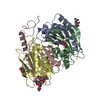
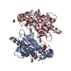
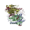
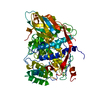
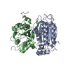
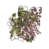
| Title | The structure of uncharacterized protein YKR043C from Saccharomyces cerevisiae. | ||||||
 Components Components | Uncharacterized protein YKR043C | ||||||
 Keywords Keywords | structural genomics / unknown function / Saccharomyces cerevisiae / PSI-2 / Protein Structure Initiative / Midwest Center for Structural Genomics / MCSG | ||||||
| Function / homology |  Function and homology information Function and homology informationsedoheptulose-bisphosphatase / sedoheptulose-bisphosphatase activity / ribose phosphate biosynthetic process / oxidoreductase activity / nucleus / cytoplasm Similarity search - Function | ||||||
| Biological species |  | ||||||
| Method |  X-RAY DIFFRACTION / SYNCHROTRON / MAD / Resolution: 1.75 Å X-RAY DIFFRACTION / SYNCHROTRON / MAD / Resolution: 1.75 Å | ||||||
 Authors Authors | Cuff, M. / Xu, X. / Cui, H. / Edwards, A. / Savchenko, A. / Joachimiak, A. / Midwest Center for Structural Genomics (MCSG) | ||||||
 Citation Citation | Journal: J.Biol.Chem. / Year: 2010 Title: Structure and activity of the metal-independent fructose-1,6-bisphosphatase YK23 from Saccharomyces cerevisiae. Authors: Kuznetsova, E. / Xu, L. / Singer, A. / Brown, G. / Dong, A. / Flick, R. / Cui, H. / Cuff, M. / Joachimiak, A. / Savchenko, A. / Yakunin, A.F. | ||||||
| History |
|
- Structure visualization
| Structure viewer | Molecule: MolmilJmol/JSmol |
|---|
- Downloads & links
-Download
| PDBx/mmCIF format | 3f3k.cif.gz | 144.1 KB | Display | PDBx/mmCIF format |
|---|---|---|---|---|
| PDB format | pdb3f3k.ent.gz | 110.9 KB | Display | PDB format |
| PDBx/mmJSON format | 3f3k.json.gz | Tree view | PDBx/mmJSON format | |
| Others |  Other downloads Other downloads |
-Validation report
| Arichive directory | https://data.pdbj.org/pub/pdb/validation_reports/f3/3f3kftp://data.pdbj.org/pub/pdb/validation_reports/f3/3f3k | HTTPS FTP |
|---|
-Related structure data


-Links
 PDBj
PDBj


- Assembly
| Deposited unit | 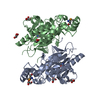
| ||||||||
|---|---|---|---|---|---|---|---|---|---|
| 1 |
| ||||||||
| Unit cell |
| ||||||||
| Details | Authors state that the biological unit is experimentally unknown. |
-Components
| #1: Protein | Mass: 30511.799 Da / Num. of mol.: 2 Source method: isolated from a genetically manipulated source Source: (gene. exp.) Strain: S288C / Gene: YKR043C / Plasmid: modified p11 / Production host:  #2: Chemical | ChemComp-GOL /   Mass: 92.094 Da / Num. of mol.: 6 / Source method: obtained synthetically / Formula: C3H8O3 Mass: 92.094 Da / Num. of mol.: 6 / Source method: obtained synthetically / Formula: C3H8O3#3: Water | ChemComp-HOH / |  Mass: 18.015 Da / Num. of mol.: 939 / Source method: isolated from a natural source / Formula: H2O Mass: 18.015 Da / Num. of mol.: 939 / Source method: isolated from a natural source / Formula: H2OHas protein modification | Y | |
|---|
-Experimental details
-Experiment
| Experiment | Method: X-RAY DIFFRACTION / Number of used crystals: 1 |
|---|
- Sample preparation
| Crystal | Density Matthews: 2.98 Å3/Da / Density % sol: 58.66 % |
|---|---|
| Crystal grow | Temperature: 293 K / Method: vapor diffusion, hanging drop / pH: 6 Details: 0.2M Li3Citrate, 16% PEG 3350, 4%MPD, 10% Glycerol, Trypsin 1/10, VAPOR DIFFUSION, HANGING DROP, temperature 293K |
-Data collection
| Diffraction | Mean temperature: 100 K | |||||||||
|---|---|---|---|---|---|---|---|---|---|---|
| Diffraction source | Source: SYNCHROTRON / Site: APS  / Beamline: 19-ID / Wavelength: 0.97948,0.97931 / Beamline: 19-ID / Wavelength: 0.97948,0.97931 | |||||||||
| Detector | Type: ADSC QUANTUM 315 / Detector: CCD / Date: Jun 29, 2008 | |||||||||
| Radiation | Monochromator: SAGITALLY FOCUSED Si(111) / Protocol: MAD / Monochromatic (M) / Laue (L): M / Scattering type: x-ray | |||||||||
| Radiation wavelength |
| |||||||||
| Reflection | Resolution: 1.75→50 Å / Num. all: 72754 / Num. obs: 72754 / % possible obs: 98.2 % / Observed criterion σ(I): -3 / Redundancy: 9.9 % / Rmerge(I) obs: 0.074 / Χ2: 1.694 / Net I/σ(I): 40.5 | |||||||||
| Reflection shell | Resolution: 1.75→1.78 Å / Redundancy: 8.9 % / Rmerge(I) obs: 0.265 / Num. unique all: 3587 / Χ2: 0.839 / % possible all: 98.4 |
-Phasing
| Phasing | Method: MAD | ||||||||||||||||||||||||||||||||||||||||||||||||||||||||||||||||||||||||||||||||||||||||||||||||||||||||||||||||||||||||||||||||||||||||||||||||||||||||||||||||||||||||||
|---|---|---|---|---|---|---|---|---|---|---|---|---|---|---|---|---|---|---|---|---|---|---|---|---|---|---|---|---|---|---|---|---|---|---|---|---|---|---|---|---|---|---|---|---|---|---|---|---|---|---|---|---|---|---|---|---|---|---|---|---|---|---|---|---|---|---|---|---|---|---|---|---|---|---|---|---|---|---|---|---|---|---|---|---|---|---|---|---|---|---|---|---|---|---|---|---|---|---|---|---|---|---|---|---|---|---|---|---|---|---|---|---|---|---|---|---|---|---|---|---|---|---|---|---|---|---|---|---|---|---|---|---|---|---|---|---|---|---|---|---|---|---|---|---|---|---|---|---|---|---|---|---|---|---|---|---|---|---|---|---|---|---|---|---|---|---|---|---|---|---|---|
| Phasing MAD | D res high: 1.75 Å / D res low: 50 Å / FOM : 0.387 / FOM acentric: 0.405 / FOM centric: 0.17 / Reflection: 72654 / Reflection acentric: 67134 / Reflection centric: 5520 | ||||||||||||||||||||||||||||||||||||||||||||||||||||||||||||||||||||||||||||||||||||||||||||||||||||||||||||||||||||||||||||||||||||||||||||||||||||||||||||||||||||||||||
| Phasing MAD set | Highest resolution: 1.75 Å / Lowest resolution: 50 Å
| ||||||||||||||||||||||||||||||||||||||||||||||||||||||||||||||||||||||||||||||||||||||||||||||||||||||||||||||||||||||||||||||||||||||||||||||||||||||||||||||||||||||||||
| Phasing MAD set shell |
| ||||||||||||||||||||||||||||||||||||||||||||||||||||||||||||||||||||||||||||||||||||||||||||||||||||||||||||||||||||||||||||||||||||||||||||||||||||||||||||||||||||||||||
| Phasing MAD set site |
| ||||||||||||||||||||||||||||||||||||||||||||||||||||||||||||||||||||||||||||||||||||||||||||||||||||||||||||||||||||||||||||||||||||||||||||||||||||||||||||||||||||||||||
| Phasing MAD shell |
| ||||||||||||||||||||||||||||||||||||||||||||||||||||||||||||||||||||||||||||||||||||||||||||||||||||||||||||||||||||||||||||||||||||||||||||||||||||||||||||||||||||||||||
| Phasing dm | Method: Solvent flattening and Histogram matching / Reflection: 72654 | ||||||||||||||||||||||||||||||||||||||||||||||||||||||||||||||||||||||||||||||||||||||||||||||||||||||||||||||||||||||||||||||||||||||||||||||||||||||||||||||||||||||||||
| Phasing dm shell |
|
- Processing
| Software |
| |||||||||||||||||||||||||||||||||||||||||||||||||||||||||||||||||||||||||||||||||||||
|---|---|---|---|---|---|---|---|---|---|---|---|---|---|---|---|---|---|---|---|---|---|---|---|---|---|---|---|---|---|---|---|---|---|---|---|---|---|---|---|---|---|---|---|---|---|---|---|---|---|---|---|---|---|---|---|---|---|---|---|---|---|---|---|---|---|---|---|---|---|---|---|---|---|---|---|---|---|---|---|---|---|---|---|---|---|---|
| Refinement | Method to determine structure: MAD / Resolution: 1.75→43.15 Å / Cor.coef. Fo:Fc: 0.975 / Cor.coef. Fo:Fc free: 0.963 / WRfactor Rfree: 0.176 / WRfactor Rwork: 0.146 / Occupancy max: 1 / Occupancy min: 0.5 / FOM work R set: 0.901 / SU B: 2.901 / SU ML: 0.043 / SU R Cruickshank DPI: 0.086 / SU Rfree: 0.086 / TLS residual ADP flag: LIKELY RESIDUAL / Cross valid method: THROUGHOUT / σ(F): 0 / ESU R: 0.085 / ESU R Free: 0.085 Stereochemistry target values: MAXIMUM LIKELIHOOD WITH PHASES Details: HYDROGENS HAVE BEEN ADDED IN THE RIDING POSITIONS U VALUES : RESIDUAL ONLY
| |||||||||||||||||||||||||||||||||||||||||||||||||||||||||||||||||||||||||||||||||||||
| Solvent computation | Ion probe radii: 0.8 Å / Shrinkage radii: 0.8 Å / VDW probe radii: 1.2 Å / Solvent model: MASK | |||||||||||||||||||||||||||||||||||||||||||||||||||||||||||||||||||||||||||||||||||||
| Displacement parameters | Biso max: 63.51 Å2 / Biso mean: 15.734 Å2 / Biso min: 5.91 Å2
| |||||||||||||||||||||||||||||||||||||||||||||||||||||||||||||||||||||||||||||||||||||
| Refinement step | Cycle: LAST / Resolution: 1.75→43.15 Å
| |||||||||||||||||||||||||||||||||||||||||||||||||||||||||||||||||||||||||||||||||||||
| Refine LS restraints |
| |||||||||||||||||||||||||||||||||||||||||||||||||||||||||||||||||||||||||||||||||||||
| LS refinement shell | Resolution: 1.75→1.795 Å / Total num. of bins used: 20
| |||||||||||||||||||||||||||||||||||||||||||||||||||||||||||||||||||||||||||||||||||||
| Refinement TLS params. | Method: refined / Refine-ID: X-RAY DIFFRACTION
| |||||||||||||||||||||||||||||||||||||||||||||||||||||||||||||||||||||||||||||||||||||
| Refinement TLS group |
|
