[English] 日本語
Yorodumi
- PDB-3f2c: DNA Polymerase PolC from Geobacillus kaustophilus complex with DN... -

+
Open data


ID or keywords:

Loading...

-
Basic information

Entry
Database: PDB / ID: 3f2c
TitleDNA Polymerase PolC from Geobacillus kaustophilus complex with DNA, dGTP and Mn
Components
  • 5'-D(*DAP*DTP*DAP*DAP*DCP*DGP*DGP*DTP*DTP*DGP*DCP*DCP*DCP*DGP*DTP*DCP*DTP*DCP*DAP*DCP*DTP*DG)-3'
  • 5'-D(*DCP*DAP*DGP*DTP*DGP*DAP*DGP*DAP*DCP*DGP*DGP*DGP*DCP*DAP*DAP*DCP*DC)-3'
  • GEOBACILLUS KAUSTOPHILUS DNA POLC
Keywords
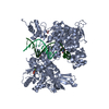
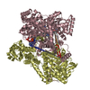
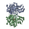
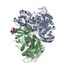
TRANSFERASE/DNA / DNA Polymerase C / DNA Polymerase III / Ternary Complex / Protein-DNA complex / Replicative polymerase / gram-positive / TRANSFERASE-DNA COMPLEX
Function / homology
Function and homology information


3'-5' exonuclease activity / DNA-templated DNA replication / DNA-directed DNA polymerase / DNA-directed DNA polymerase activity / DNA binding / metal ion binding / cytoplasm
Similarity search - Function
PolC, middle finger domain / DNA polymerase; domain 1 - #870 / glyoxalase-related enzyme like fold - #20 / Helix Hairpins - #1510 / Insulin-like, subunit E - #10 / DNA polymerase III PolC-like, N-terminal domain II / DNA polymerase III PolC-type, N-terminal domain I / PolC, middle finger subdomain superfamily / DNA polymerase III polC-type N-terminus II / DNA polymerase III polC-type N-terminus I ...PolC, middle finger domain / DNA polymerase; domain 1 - #870 / glyoxalase-related enzyme like fold - #20 / Helix Hairpins - #1510 / Insulin-like, subunit E - #10 / DNA polymerase III PolC-like, N-terminal domain II / DNA polymerase III PolC-type, N-terminal domain I / PolC, middle finger subdomain superfamily / DNA polymerase III polC-type N-terminus II / DNA polymerase III polC-type N-terminus I / glyoxalase-related enzyme like fold / Insulin-like, subunit E / DNA polymerase III, alpha subunit, Gram-positive type / DNA polymerase III epsilon subunit, exonuclease domain / DNA polymerase III, alpha subunit / Bacterial DNA polymerase III, alpha subunit, NTPase domain / DNA polymerase, helix-hairpin-helix motif / DNA polymerase III alpha subunit finger domain / Bacterial DNA polymerase III alpha NTPase domain / Helix-hairpin-helix motif / Bacterial DNA polymerase III alpha subunit finger domain / PHP domain / PHP domain / Polymerase/histidinol phosphatase, N-terminal / DNA polymerase alpha chain like domain / Exonuclease / Exonuclease, RNase T/DNA polymerase III / EXOIII / OB-fold nucleic acid binding domain, AA-tRNA synthetase-type / OB-fold nucleic acid binding domain / Helix Hairpins / Metal-dependent hydrolases / Nucleic acid-binding proteins / Helix non-globular / Special / DNA polymerase; domain 1 / OB fold (Dihydrolipoamide Acetyltransferase, E2P) / Ribonuclease H superfamily / Ribonuclease H-like superfamily / Nucleic acid-binding, OB-fold / TIM Barrel / Alpha-Beta Barrel / Beta Barrel / 2-Layer Sandwich / Orthogonal Bundle / Mainly Beta / Mainly Alpha / Alpha Beta
Similarity search - Domain/homology
2'-DEOXYGUANOSINE-5'-TRIPHOSPHATE / : / DNA / DNA (> 10) / DNA polymerase III PolC-type
Similarity search - Component
Biological speciesGeobacillus kaustophilus (bacteria)
MethodX-RAY DIFFRACTION / SYNCHROTRON / MOLECULAR REPLACEMENT / Resolution: 2.5 Å
AuthorsDavies, D.R. / Evans, R.J. / Bullard, J.M. / Christensen, J. / Green, L.S. / Guiles, J.W. / Ribble, W.K. / Janjic, N. / Jarvis, T.C.
CitationJournal: Proc.Natl.Acad.Sci.USA / Year: 2008
Title: Structure of PolC reveals unique DNA binding and fidelity determinants.
Authors: Evans, R.J. / Davies, D.R. / Bullard, J.M. / Christensen, J. / Green, L.S. / Guiles, J.W. / Pata, J.D. / Ribble, W.K. / Janjic, N. / Jarvis, T.C.
History
DepositionOct 29, 2008Deposition site: RCSB / Processing site: RCSB
Revision 1.0Jan 20, 2009Provider: repository / Type: Initial release
Revision 1.1Jul 13, 2011Group: Advisory / Refinement description / Version format compliance
Revision 1.2Jul 26, 2017Group: Refinement description / Source and taxonomy / Category: entity_src_gen / software
Revision 1.3Sep 6, 2023Group: Data collection / Database references ...Data collection / Database references / Derived calculations / Refinement description
Category: chem_comp_atom / chem_comp_bond ...chem_comp_atom / chem_comp_bond / database_2 / pdbx_initial_refinement_model / pdbx_struct_conn_angle / struct_conn / struct_ref_seq_dif / struct_site
Item: _database_2.pdbx_DOI / _database_2.pdbx_database_accession ..._database_2.pdbx_DOI / _database_2.pdbx_database_accession / _pdbx_struct_conn_angle.ptnr1_auth_comp_id / _pdbx_struct_conn_angle.ptnr1_auth_seq_id / _pdbx_struct_conn_angle.ptnr1_label_asym_id / _pdbx_struct_conn_angle.ptnr1_label_atom_id / _pdbx_struct_conn_angle.ptnr1_label_comp_id / _pdbx_struct_conn_angle.ptnr1_label_seq_id / _pdbx_struct_conn_angle.ptnr2_auth_comp_id / _pdbx_struct_conn_angle.ptnr2_auth_seq_id / _pdbx_struct_conn_angle.ptnr2_label_asym_id / _pdbx_struct_conn_angle.ptnr2_label_atom_id / _pdbx_struct_conn_angle.ptnr2_label_comp_id / _pdbx_struct_conn_angle.ptnr3_auth_comp_id / _pdbx_struct_conn_angle.ptnr3_auth_seq_id / _pdbx_struct_conn_angle.ptnr3_label_asym_id / _pdbx_struct_conn_angle.ptnr3_label_atom_id / _pdbx_struct_conn_angle.ptnr3_label_comp_id / _pdbx_struct_conn_angle.ptnr3_label_seq_id / _pdbx_struct_conn_angle.value / _struct_conn.pdbx_dist_value / _struct_conn.ptnr1_auth_comp_id / _struct_conn.ptnr1_auth_seq_id / _struct_conn.ptnr1_label_asym_id / _struct_conn.ptnr1_label_atom_id / _struct_conn.ptnr1_label_comp_id / _struct_conn.ptnr1_label_seq_id / _struct_conn.ptnr2_auth_comp_id / _struct_conn.ptnr2_auth_seq_id / _struct_conn.ptnr2_label_asym_id / _struct_conn.ptnr2_label_atom_id / _struct_conn.ptnr2_label_comp_id / _struct_conn.ptnr2_label_seq_id / _struct_ref_seq_dif.details / _struct_site.pdbx_auth_asym_id / _struct_site.pdbx_auth_comp_id / _struct_site.pdbx_auth_seq_id

-
Structure visualization

Structure viewerMolecule:
MolmilJmol/JSmol

Downloads & links

-
Assembly

Deposited unit
A: GEOBACILLUS KAUSTOPHILUS DNA POLC
P: 5'-D(*DCP*DAP*DGP*DTP*DGP*DAP*DGP*DAP*DCP*DGP*DGP*DGP*DCP*DAP*DAP*DCP*DC)-3'
T: 5'-D(*DAP*DTP*DAP*DAP*DCP*DGP*DGP*DTP*DTP*DGP*DCP*DCP*DCP*DGP*DTP*DCP*DTP*DCP*DAP*DCP*DTP*DG)-3'
hetero molecules


Theoretical massNumber of molelcules
Total (without water)130,0329
Polymers129,1983
Non-polymers8336
Water2,792155
1


  • Idetical with deposited unit
  • defined by author&software
TypeNameSymmetry operationNumber
identity operation1_555x,y,z1
Buried area6420 Å2
ΔGint-72 kcal/mol
Surface area45170 Å2
MethodPISA
Unit cell
Length a, b, c (Å)115.889, 141.185, 184.740
Angle α, β, γ (deg.)90.00, 90.00, 90.00
Int Tables number20
Space group name H-MC2221
DetailsBIOLOGICAL UNIT IS THE SAME AS ASYMMETRIC UNIT

-
Components

-
Protein , 1 types, 1 molecules A

#1: Protein GEOBACILLUS KAUSTOPHILUS DNA POLC


Mass: 117248.508 Da / Num. of mol.: 1 / Fragment: GKAPOLC, DELTA 1-227, DELTA 412-617
Source method: isolated from a genetically manipulated source
Source: (gene. exp.) Geobacillus kaustophilus (bacteria) / Production host: Escherichia coli (E. coli) / References: UniProt: Q5L0J3, DNA-directed DNA polymerase

-
DNA chain , 2 types, 2 molecules PT

#2: DNA chain 5'-D(*DCP*DAP*DGP*DTP*DGP*DAP*DGP*DAP*DCP*DGP*DGP*DGP*DCP*DAP*DAP*DCP*DC)-3'


Mass: 5246.413 Da / Num. of mol.: 1 / Source method: obtained synthetically / Details: Primer strand is synthetic oligonucleotide
#3: DNA chain 5'-D(*DAP*DTP*DAP*DAP*DCP*DGP*DGP*DTP*DTP*DGP*DCP*DCP*DCP*DGP*DTP*DCP*DTP*DCP*DAP*DCP*DTP*DG)-3'


Mass: 6703.328 Da / Num. of mol.: 1 / Source method: obtained synthetically / Details: Template strand is synthetic oligonucleotide

-
Non-polymers , 5 types, 161 molecules

#4: Chemical ChemComp-MN / MANGANESE (II) ION


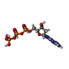
Mass: 54.938 Da / Num. of mol.: 3 / Source method: obtained synthetically / Formula: Mn
#5: Chemical ChemComp-ZN / ZINC ION


Mass: 65.409 Da / Num. of mol.: 1 / Source method: obtained synthetically / Formula: Zn
#6: Chemical ChemComp-SO4 / SULFATE ION


Mass: 96.063 Da / Num. of mol.: 1 / Source method: obtained synthetically / Formula: SO4
#7: Chemical ChemComp-DGT / 2'-DEOXYGUANOSINE-5'-TRIPHOSPHATE


Mass: 507.181 Da / Num. of mol.: 1 / Source method: obtained synthetically / Formula: C10H16N5O13P3
#8: Water ChemComp-HOH / water


Mass: 18.015 Da / Num. of mol.: 155 / Source method: isolated from a natural source / Formula: H2O

-
Experimental details

-
Experiment

ExperimentMethod: X-RAY DIFFRACTION / Number of used crystals: 1

-
Sample preparation

CrystalDensity Matthews: 2.92 Å3/Da / Density % sol: 57.94 %
Crystal growTemperature: 289 K / Method: vapor diffusion, sitting drop / pH: 5.3
Details: 20% PEG 2000 MME, 0.1M PHOSPHATE CITRATE, 200MM LITHIUM SULFATE, 2 mM MnCl2, PH 5.3, VAPOR DIFFUSION , pH 5.30, VAPOR DIFFUSION, SITTING DROP, temperature 289K
Components of the solutions
IDNameCrystal-IDSol-ID
1PEG 2000 MME11
2PHOSPHATE CITRATE11
3LITHIUM SULFATE11
4MnCl211
5PHOSPHATE CITRATE12
6LITHIUM SULFATE12
7PEG 2000 MME12
8MnCl212

-
Data collection

DiffractionMean temperature: 100 K
Diffraction sourceSource: SYNCHROTRON / Site: ALS / Beamline: 5.0.1 / Wavelength: 0.97 / Wavelength: 0.999887 Å
DetectorDetector: CCD / Date: Apr 17, 2008
RadiationProtocol: SINGLE WAVELENGTH / Monochromatic (M) / Laue (L): M / Scattering type: x-ray
Radiation wavelength
IDWavelength (Å)Relative weight
10.971
20.9998871
ReflectionResolution: 2.5→43.6 Å / Num. obs: 51829 / % possible obs: 99.1 % / Redundancy: 5.9 % / Rmerge(I) obs: 0.09 / Net I/σ(I): 10
Reflection shellResolution: 2.5→2.59 Å / Redundancy: 3.9 % / Rmerge(I) obs: 0.589 / % possible all: 91.7

-
Processing

Software
NameVersionClassification
HKL-2000data collection
MOLREPphasing
REFMAC5.4.0069refinement
HKL-2000data reduction
HKL-2000data scaling
RefinementMethod to determine structure: MOLECULAR REPLACEMENT
Starting model: PDB entry 3F2B
Resolution: 2.5→43.6 Å / Cor.coef. Fo:Fc: 0.937 / Cor.coef. Fo:Fc free: 0.912 / SU B: 17.736 / SU ML: 0.203 / TLS residual ADP flag: LIKELY RESIDUAL / Cross valid method: THROUGHOUT / σ(F): 0 / ESU R: 0.413 / ESU R Free: 0.273 / Stereochemistry target values: MAXIMUM LIKELIHOOD / Details: HYDROGENS HAVE BEEN ADDED IN THE RIDING POSITIONS
RfactorNum. reflection% reflectionSelection details
Rfree0.255 2641 5.1 %RANDOM
Rwork0.21 ---
obs0.213 51791 --
Solvent computationIon probe radii: 0.8 Å / Shrinkage radii: 0.8 Å / VDW probe radii: 1.2 Å / Solvent model: MASK
Displacement parametersBiso mean: 39.53 Å2
Baniso -1Baniso -2Baniso -3
1--1.82 Å20 Å20 Å2
2--2.62 Å20 Å2
3----0.8 Å2
Refinement stepCycle: LAST / Resolution: 2.5→43.6 Å
ProteinNucleic acidLigandSolventTotal
Num. atoms7796 631 40 155 8622
Refine LS restraints
Refine-IDTypeDev idealDev ideal targetNumber
X-RAY DIFFRACTIONr_bond_refined_d0.010.0228704
X-RAY DIFFRACTIONr_bond_other_d
X-RAY DIFFRACTIONr_angle_refined_deg1.4522.06711920
X-RAY DIFFRACTIONr_angle_other_deg
X-RAY DIFFRACTIONr_dihedral_angle_1_deg5.8655989
X-RAY DIFFRACTIONr_dihedral_angle_2_deg38.35124.294361
X-RAY DIFFRACTIONr_dihedral_angle_3_deg20.096151387
X-RAY DIFFRACTIONr_dihedral_angle_4_deg20.8621546
X-RAY DIFFRACTIONr_chiral_restr0.0940.21333
X-RAY DIFFRACTIONr_gen_planes_refined0.0060.0216338
X-RAY DIFFRACTIONr_gen_planes_other
X-RAY DIFFRACTIONr_nbd_refined
X-RAY DIFFRACTIONr_nbd_other
X-RAY DIFFRACTIONr_nbtor_refined
X-RAY DIFFRACTIONr_nbtor_other
X-RAY DIFFRACTIONr_xyhbond_nbd_refined
X-RAY DIFFRACTIONr_xyhbond_nbd_other
X-RAY DIFFRACTIONr_metal_ion_refined
X-RAY DIFFRACTIONr_metal_ion_other
X-RAY DIFFRACTIONr_symmetry_vdw_refined
X-RAY DIFFRACTIONr_symmetry_vdw_other
X-RAY DIFFRACTIONr_symmetry_hbond_refined
X-RAY DIFFRACTIONr_symmetry_hbond_other
X-RAY DIFFRACTIONr_symmetry_metal_ion_refined
X-RAY DIFFRACTIONr_symmetry_metal_ion_other
X-RAY DIFFRACTIONr_mcbond_it0.3911.54940
X-RAY DIFFRACTIONr_mcbond_other
X-RAY DIFFRACTIONr_mcangle_it0.77227993
X-RAY DIFFRACTIONr_scbond_it1.25933764
X-RAY DIFFRACTIONr_scangle_it2.0794.53927
X-RAY DIFFRACTIONr_rigid_bond_restr
X-RAY DIFFRACTIONr_sphericity_free
X-RAY DIFFRACTIONr_sphericity_bonded
LS refinement shellResolution: 2.5→2.57 Å / Total num. of bins used: 20
RfactorNum. reflection% reflection
Rfree0.364 166 -
Rwork0.323 3189 -
obs--87.28 %
Refinement TLS params.

Method: refined / Refine-ID: X-RAY DIFFRACTION

IDL112)L122)L132)L222)L232)L332)S11 (Å °)S12 (Å °)S13 (Å °)S21 (Å °)S22 (Å °)S23 (Å °)S31 (Å °)S32 (Å °)S33 (Å °)T112)T122)T132)T222)T232)T332)Origin x (Å)Origin y (Å)Origin z (Å)
16.93971.8614-1.68343.4265-2.20344.35380.0477-0.13720.04710.19470.1070.226-0.37880.0223-0.1547-0.09390.03820.0149-0.12440.0636-0.15738.208411.986716.2396
20.5268-0.29960.62612.5295-0.1013.074-0.1605-0.030.17170.1650.3536-0.0528-0.6574-0.4296-0.19310.04770.19020.0805-0.04350.0066-0.11638.3642-4.475346.4072
31.7111-0.012-0.77493.64820.28082.8538-0.0727-0.2165-0.11410.440.24420.3224-0.3418-1.0216-0.1716-0.09140.31220.11070.28180.105-0.194827.8924-12.428157.116
40.60960.1771.58941.0290.40398.2219-0.2881-0.50170.08210.37590.50050.2951-0.824-1.7698-0.21240.14470.5460.15610.33690.1148-0.092123.63641.418848.9645
50.7698-1.0055-0.1542.7929-0.40052.3983-0.2083-0.32670.16790.50980.5394-0.3862-0.7219-0.4012-0.3310.10360.2770.0434-0.01550.0064-0.191938.6683-1.178353.3823
61.4144-0.2406-0.23271.293-0.12872.81990.0890.0163-0.1739-0.0492-0.032-0.12850.56380.1901-0.0569-0.0101-0.0006-0.034-0.1509-0.004-0.051255.6121-44.676825.9865
70.24280.02220.01150.7389-0.10670.37340.0188-0.0146-0.0782-0.01850.16870.14990.0546-0.2712-0.1875-0.1116-0.01960.01690.04990.0554-0.068334.3961-24.537831.5109
81.94590.65350.53080.5855-0.13641.2568-0.00050.1668-0.008-0.14060.0461-0.0466-0.06990.082-0.0456-0.0558-0.04080.0233-0.078-0.0159-0.094451.4769-28.60959.8977
94.67772.34031.50585.359-1.06523.8951-0.03770.34780.5815-0.1575-0.01060.448-0.3113-0.41350.0483-0.0996-0.0641-0.0104-0.01480.0757-0.093125.5823-30.38551.1107
108.07542.7721-1.21530.9586-0.39090.28210.0988-0.476-0.2170.2034-0.187-0.0248-0.0115-0.02720.0882-0.0503-0.1044-0.02740.05330.0172-0.08669.0143-45.274212.4399
111.6546-0.808-0.48131.26051.27041.378-0.1848-0.0209-0.1744-0.12-0.19990.48120.3065-0.51380.3847-0.1237-0.0816-0.04850.1708-0.00620.144723.5739-24.458519.952
Refinement TLS group
IDRefine-IDRefine TLS-IDAuth asym-IDAuth seq-ID
1X-RAY DIFFRACTION1A233 - 331
2X-RAY DIFFRACTION2A332 - 411
3X-RAY DIFFRACTION3A619 - 708
4X-RAY DIFFRACTION4A716 - 772
5X-RAY DIFFRACTION5A773 - 825
6X-RAY DIFFRACTION6A826 - 996
7X-RAY DIFFRACTION7A997 - 1122
8X-RAY DIFFRACTION8A1123 - 1307
9X-RAY DIFFRACTION9A1308 - 1359
10X-RAY DIFFRACTION10A1360 - 1444
11X-RAY DIFFRACTION11P5 - 17
12X-RAY DIFFRACTION11T1 - 18

+
About Yorodumi

-
News

-
Feb 9, 2022. New format data for meta-information of EMDB entries

New format data for meta-information of EMDB entries

  • Version 3 of the EMDB header file is now the official format.
  • The previous official version 1.9 will be removed from the archive.

Related info.:EMDB header

External links:wwPDB to switch to version 3 of the EMDB data model

-
Aug 12, 2020. Covid-19 info

Covid-19 info

URL: https://pdbjlvh1.pdbj.org/emnavi/covid19.php

New page: Covid-19 featured information page in EM Navigator.

Related info.:Covid-19 info / Mar 5, 2020. Novel coronavirus structure data

+
Mar 5, 2020. Novel coronavirus structure data

Novel coronavirus structure data

Related info.:Yorodumi Speices / Aug 12, 2020. Covid-19 info

External links:COVID-19 featured content - PDBj / Molecule of the Month (242):Coronavirus Proteases

+
Jan 31, 2019. EMDB accession codes are about to change! (news from PDBe EMDB page)

EMDB accession codes are about to change! (news from PDBe EMDB page)

  • The allocation of 4 digits for EMDB accession codes will soon come to an end. Whilst these codes will remain in use, new EMDB accession codes will include an additional digit and will expand incrementally as the available range of codes is exhausted. The current 4-digit format prefixed with “EMD-” (i.e. EMD-XXXX) will advance to a 5-digit format (i.e. EMD-XXXXX), and so on. It is currently estimated that the 4-digit codes will be depleted around Spring 2019, at which point the 5-digit format will come into force.
  • The EM Navigator/Yorodumi systems omit the EMD- prefix.

Related info.:Q: What is EMD? / ID/Accession-code notation in Yorodumi/EM Navigator

External links:EMDB Accession Codes are Changing Soon! / Contact to PDBj

+
Jul 12, 2017. Major update of PDB

Major update of PDB

  • wwPDB released updated PDB data conforming to the new PDBx/mmCIF dictionary.
  • This is a major update changing the version number from 4 to 5, and with Remediation, in which all the entries are updated.
  • In this update, many items about electron microscopy experimental information are reorganized (e.g. em_software).
  • Now, EM Navigator and Yorodumi are based on the updated data.

External links:wwPDB Remediation / Enriched Model Files Conforming to OneDep Data Standards Now Available in the PDB FTP Archive

-
Yorodumi

Thousand views of thousand structures

  • Yorodumi is a browser for structure data from EMDB, PDB, SASBDB, etc.
  • This page is also the successor to EM Navigator detail page, and also detail information page/front-end page for Omokage search.
  • The word "yorodu" (or yorozu) is an old Japanese word meaning "ten thousand". "mi" (miru) is to see.

Related info.:EMDB / PDB / SASBDB / Comparison of 3 databanks / Yorodumi Search / Aug 31, 2016. New EM Navigator & Yorodumi / Yorodumi Papers / Jmol/JSmol / Function and homology information / Changes in new EM Navigator and Yorodumi

Read more