- PDB-3ez0: Crystal structure of protein of unknown function with ferritin-li... -
+
Open data
ID or keywords:
Loading...
-
Basic information
Entry
Database: PDB / ID: 3ez0
Title
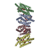
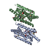
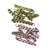
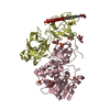
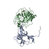
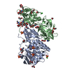
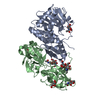
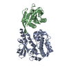
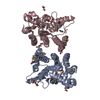
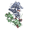
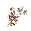
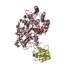
Crystal structure of protein of unknown function with ferritin-like fold (YP_832262.1) from Arthrobacter sp. FB24 at 2.33 A resolution
Components
uncharacterized protein with ferritin-like fold
Keywords
structural genomics / unknown function / YP_832262.1 / protein of unknown function with ferritin-like fold / Joint Center for Structural Genomics / JCSG / Protein Structure Initiative / PSI-2
#228 - Dec 2018 Directed Evolution of Enzymes similarity (1)
-
Assembly
Deposited unit
A: uncharacterized protein with ferritin-like fold B: uncharacterized protein with ferritin-like fold C: uncharacterized protein with ferritin-like fold D: uncharacterized protein with ferritin-like fold hetero molecules
 Movie
Movie Controller

 Yorodumi
 Open data
 Basic information
 Components
 Keywords
 Function and homology information
 Arthrobacter sp. FB24 (bacteria)
 X-RAY DIFFRACTION /
  Authors
 Citation
 Structure visualization
 Downloads & links
 Other downloads
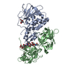
 PDBj
PDBj


 Assembly