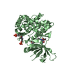
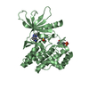
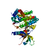
Entry Database : PDB / ID : 3eygTitle Crystal structures of JAK1 and JAK2 inhibitor complexes Tyrosine-protein kinase Keywords / / / / / / / Function / homology Function Domain/homology Component
/ / / / / / / / / / / / / / / / / / / / / / / / / / / / / / / / / / / / / / / / / / / / / / / / / / / / / / / / / / / / / / / / / / / / / / / / / / / / / / / / / / / / / / / / / / / / / / / / / / / / / / / / / / / / / / / / / / / / / / / / / / / / / / / / / Biological species Homo sapiens (human)Method / / Resolution : 1.9 Å Authors Williams, N.K. / Bamert, R.S. / Patell, O. / Wang, C. / Walden, P.M. / Fantino, E. Journal : J.Mol.Biol. / Year : 2009Title : Dissecting specificity in the Janus kinases: the structures of JAK-specific inhibitors complexed to the JAK1 and JAK2 protein tyrosine kinase domains.Authors : Williams, N.K. / Bamert, R.S. / Patel, O. / Wang, C. / Walden, P.M. / Wilks, A.F. / Fantino, E. / Rossjohn, J. / Lucet, I.S. History Deposition Oct 20, 2008 Deposition site / Processing site Revision 1.0 Feb 3, 2009 Provider / Type Revision 1.1 Jul 13, 2011 Group Revision 1.2 Dec 27, 2023 Group / Database references / Derived calculationsCategory chem_comp_atom / chem_comp_bond ... chem_comp_atom / chem_comp_bond / database_2 / struct_conn / struct_site Item _database_2.pdbx_DOI / _database_2.pdbx_database_accession ... _database_2.pdbx_DOI / _database_2.pdbx_database_accession / _struct_conn.pdbx_leaving_atom_flag / _struct_site.pdbx_auth_asym_id / _struct_site.pdbx_auth_comp_id / _struct_site.pdbx_auth_seq_id Revision 1.3 Oct 30, 2024 Group / Category / pdbx_modification_feature
Show all Show less
 Movie
Movie Controller


 Open data
 Basic information
 Components
 Keywords
 Function and homology information
 Homo sapiens (human)
 X-RAY DIFFRACTION /
  Authors
 Citation
 Structure visualization
 Downloads & links
 Other downloads
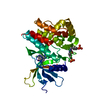


 PDBj
PDBj


 Assembly


 Mass: 312.370 Da / Num. of mol.: 1 / Source method: obtained synthetically / Formula: C16H20N6O / Comment: medication, inhibitor*YM
 Mass: 18.015 Da / Num. of mol.: 265 / Source method: isolated from a natural source / Formula: H2O
 Sample preparation
 / Beamline: MX1
 Processing