 Movie
Movie Controller

[English] 日本語
 Yorodumi
- PDB-3exh: Crystal structure of the pyruvate dehydrogenase (E1p) component o... -
+ Open data

- Basic information
| Entry | Database: PDB / ID: 3exh | ||||||
|---|---|---|---|---|---|---|---|
| Title | Crystal structure of the pyruvate dehydrogenase (E1p) component of human pyruvate dehydrogenase complex | ||||||
 Components Components |
| ||||||
 Keywords Keywords | OXIDOREDUCTASE / heterotetramer / thiamine diphosphate-dependent enzyme / Disease mutation / Glycolysis / Leigh syndrome / Mitochondrion / Phosphoprotein / Alternative splicing / Polymorphism / Pyruvate / Thiamine pyrophosphate / Transit peptide | ||||||
| Function / homology |  Function and homology information Function and homology informationpyruvate dehydrogenase (acetyl-transferring) / pyruvate dehydrogenase (acetyl-transferring) activity / PDH complex synthesizes acetyl-CoA from PYR / pyruvate decarboxylation to acetyl-CoA / Regulation of pyruvate dehydrogenase (PDH) complex / pyruvate dehydrogenase complex / Signaling by Retinoic Acid / tricarboxylic acid cycle / Mitochondrial protein degradation / glucose metabolic process ...pyruvate dehydrogenase (acetyl-transferring) / pyruvate dehydrogenase (acetyl-transferring) activity / PDH complex synthesizes acetyl-CoA from PYR / pyruvate decarboxylation to acetyl-CoA / Regulation of pyruvate dehydrogenase (PDH) complex / pyruvate dehydrogenase complex / Signaling by Retinoic Acid / tricarboxylic acid cycle / Mitochondrial protein degradation / glucose metabolic process / mitochondrial matrix / nucleolus / mitochondrion / nucleoplasm / metal ion binding / nucleus Similarity search - Function | ||||||
| Biological species |  Homo sapiens (human) Homo sapiens (human) | ||||||
| Method |  X-RAY DIFFRACTION / Resolution: 2.444 Å X-RAY DIFFRACTION / Resolution: 2.444 Å | ||||||
 Authors Authors | Kato, M. / Wynn, R.M. / Chuang, J.L. / Tso, S.-C. / Machius, M. / Li, J. / Chuang, D.T. | ||||||
 Citation Citation | Journal: Structure / Year: 2008 Title: Structural basis for inactivation of the human pyruvate dehydrogenase complex by phosphorylation: role of disordered phosphorylation loops. Authors: Kato, M. / Wynn, R.M. / Chuang, J.L. / Tso, S.C. / Machius, M. / Li, J. / Chuang, D.T. | ||||||
| History |
|
- Structure visualization
| Structure viewer | Molecule: MolmilJmol/JSmol |
|---|
- Downloads & links
-Download
| PDBx/mmCIF format | 3exh.cif.gz | 548.5 KB | Display | PDBx/mmCIF format |
|---|---|---|---|---|
| PDB format | pdb3exh.ent.gz | 441.5 KB | Display | PDB format |
| PDBx/mmJSON format | 3exh.json.gz | Tree view | PDBx/mmJSON format | |
| Others |  Other downloads Other downloads |
-Validation report
| Arichive directory | https://data.pdbj.org/pub/pdb/validation_reports/ex/3exhftp://data.pdbj.org/pub/pdb/validation_reports/ex/3exh | HTTPS FTP |
|---|
-Related structure data


-Links
 PDBj
PDBj


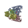
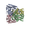
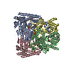
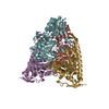
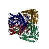
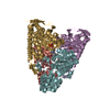
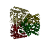
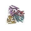
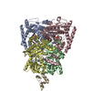
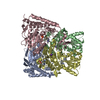
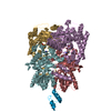
- Assembly
| Deposited unit | 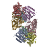
| |||||||||||||||||||||||||||||||||||||||||||||||||||||||||||||||||||||||||||||||||||||||||||||||||||||||||||||||||||||||||||||||||||||||||||||||||||||
|---|---|---|---|---|---|---|---|---|---|---|---|---|---|---|---|---|---|---|---|---|---|---|---|---|---|---|---|---|---|---|---|---|---|---|---|---|---|---|---|---|---|---|---|---|---|---|---|---|---|---|---|---|---|---|---|---|---|---|---|---|---|---|---|---|---|---|---|---|---|---|---|---|---|---|---|---|---|---|---|---|---|---|---|---|---|---|---|---|---|---|---|---|---|---|---|---|---|---|---|---|---|---|---|---|---|---|---|---|---|---|---|---|---|---|---|---|---|---|---|---|---|---|---|---|---|---|---|---|---|---|---|---|---|---|---|---|---|---|---|---|---|---|---|---|---|---|---|---|---|---|
| 1 | 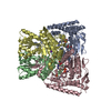
| |||||||||||||||||||||||||||||||||||||||||||||||||||||||||||||||||||||||||||||||||||||||||||||||||||||||||||||||||||||||||||||||||||||||||||||||||||||
| 2 | 
| |||||||||||||||||||||||||||||||||||||||||||||||||||||||||||||||||||||||||||||||||||||||||||||||||||||||||||||||||||||||||||||||||||||||||||||||||||||
| Unit cell |
| |||||||||||||||||||||||||||||||||||||||||||||||||||||||||||||||||||||||||||||||||||||||||||||||||||||||||||||||||||||||||||||||||||||||||||||||||||||
| Noncrystallographic symmetry (NCS) | NCS domain:
NCS domain segments: Refine code: 5
NCS ensembles :
|
-Components
-Pyruvate dehydrogenase E1 component subunit alpha, somatic form, ... , 2 types, 4 molecules AECG
| #1: Protein | Mass: 42552.516 Da / Num. of mol.: 2 / Fragment: E1p-alpha / Mutation: S203A,S271A Source method: isolated from a genetically manipulated source Details: S203A/S271A with bound Mn-ThDP / Source: (gene. exp.) Homo sapiens (human) / Gene: PDHA1, PHE1A / Production host:  References: UniProt: P08559, pyruvate dehydrogenase (acetyl-transferring) #3: Protein | Mass: 42632.492 Da / Num. of mol.: 2 / Fragment: E1p-alpha / Mutation: S203A,S271A Source method: isolated from a genetically manipulated source Details: phosphorylated S264, S203A/S271A with bound Mn-ThDP Source: (gene. exp.) Homo sapiens (human) / Gene: PDHA1, PHE1A / Production host: References: UniProt: P08559, pyruvate dehydrogenase (acetyl-transferring) |
|---|
-Protein , 1 types, 4 molecules BDFH
| #2: Protein | Mass: 35941.316 Da / Num. of mol.: 4 / Fragment: E1p-beta Source method: isolated from a genetically manipulated source Source: (gene. exp.) Homo sapiens (human) / Gene: PDHB, PHE1B / Production host: References: UniProt: P11177, pyruvate dehydrogenase (acetyl-transferring) |
|---|
-Non-polymers , 5 types, 809 molecules 


| #4: Chemical | ChemComp-MN /  Mass: 54.938 Da / Num. of mol.: 4 / Source method: obtained synthetically / Formula: Mn Mass: 54.938 Da / Num. of mol.: 4 / Source method: obtained synthetically / Formula: Mn#5: Chemical | ChemComp-K /  Mass: 39.098 Da / Num. of mol.: 4 / Source method: obtained synthetically / Formula: K Mass: 39.098 Da / Num. of mol.: 4 / Source method: obtained synthetically / Formula: K#6: Chemical | ChemComp-TPP /  Mass: 425.314 Da / Num. of mol.: 4 / Source method: obtained synthetically / Formula: C12H19N4O7P2S Mass: 425.314 Da / Num. of mol.: 4 / Source method: obtained synthetically / Formula: C12H19N4O7P2S#7: Chemical | ChemComp-GOL /  Mass: 92.094 Da / Num. of mol.: 5 / Source method: obtained synthetically / Formula: C3H8O3 Mass: 92.094 Da / Num. of mol.: 5 / Source method: obtained synthetically / Formula: C3H8O3#8: Water | ChemComp-HOH / | Mass: 18.015 Da / Num. of mol.: 792 / Source method: isolated from a natural source / Formula: H2O |
|---|
-Details
| Has protein modification | Y |
|---|
-Experimental details
-Experiment
| Experiment | Method: X-RAY DIFFRACTION / Number of used crystals: 1 |
|---|
- Sample preparation
| Crystal | Density Matthews: 2.76 Å3/Da / Density % sol: 55.51 % |
|---|---|
| Crystal grow | Temperature: 298 K / Method: vapor diffusion, hanging drop / pH: 6.5 Details: 12% PEG 8000, 140mM NaOAC, 50mM BisTris pH6.5, 10% glucose, 50mM Taurine, 8mM MgCl2, and 10mM TCEP, VAPOR DIFFUSION, HANGING DROP, temperature 298K |
-Data collection
| Diffraction | Mean temperature: 100 K | |||||||||||||||||||||||||||||||||||||||||||||||||||||||||||||||||||||||||||||||||||||||||||||||||||||||||||||||||||||||||||||||||||||||||||||||||||
|---|---|---|---|---|---|---|---|---|---|---|---|---|---|---|---|---|---|---|---|---|---|---|---|---|---|---|---|---|---|---|---|---|---|---|---|---|---|---|---|---|---|---|---|---|---|---|---|---|---|---|---|---|---|---|---|---|---|---|---|---|---|---|---|---|---|---|---|---|---|---|---|---|---|---|---|---|---|---|---|---|---|---|---|---|---|---|---|---|---|---|---|---|---|---|---|---|---|---|---|---|---|---|---|---|---|---|---|---|---|---|---|---|---|---|---|---|---|---|---|---|---|---|---|---|---|---|---|---|---|---|---|---|---|---|---|---|---|---|---|---|---|---|---|---|---|---|---|---|
| Diffraction source | Source: ROTATING ANODE / Type: RIGAKU FR-D / Wavelength: 1.5418 Å | |||||||||||||||||||||||||||||||||||||||||||||||||||||||||||||||||||||||||||||||||||||||||||||||||||||||||||||||||||||||||||||||||||||||||||||||||||
| Detector | Type: RIGAKU RAXIS IV / Detector: IMAGE PLATE / Date: May 12, 2008 | |||||||||||||||||||||||||||||||||||||||||||||||||||||||||||||||||||||||||||||||||||||||||||||||||||||||||||||||||||||||||||||||||||||||||||||||||||
| Radiation | Protocol: SINGLE WAVELENGTH / Monochromatic (M) / Laue (L): M / Scattering type: x-ray | |||||||||||||||||||||||||||||||||||||||||||||||||||||||||||||||||||||||||||||||||||||||||||||||||||||||||||||||||||||||||||||||||||||||||||||||||||
| Radiation wavelength | Wavelength: 1.5418 Å / Relative weight: 1 | |||||||||||||||||||||||||||||||||||||||||||||||||||||||||||||||||||||||||||||||||||||||||||||||||||||||||||||||||||||||||||||||||||||||||||||||||||
| Reflection | Resolution: 2.444→50 Å / Num. obs: 123888 / % possible obs: 98.1 % / Redundancy: 6.7 % / Rmerge(I) obs: 0.155 / Χ2: 1.296 / Net I/σ(I): 14.158 | |||||||||||||||||||||||||||||||||||||||||||||||||||||||||||||||||||||||||||||||||||||||||||||||||||||||||||||||||||||||||||||||||||||||||||||||||||
| Reflection shell |
|
- Processing
| Software |
| ||||||||||||||||||||||||||||||||||||||||||||||||||||||||||||||||||||||||||||||||||||||||||||||||||||||||||||||||||||||||||||||||||||||||||||||||||||||||||||||||||||||||||
|---|---|---|---|---|---|---|---|---|---|---|---|---|---|---|---|---|---|---|---|---|---|---|---|---|---|---|---|---|---|---|---|---|---|---|---|---|---|---|---|---|---|---|---|---|---|---|---|---|---|---|---|---|---|---|---|---|---|---|---|---|---|---|---|---|---|---|---|---|---|---|---|---|---|---|---|---|---|---|---|---|---|---|---|---|---|---|---|---|---|---|---|---|---|---|---|---|---|---|---|---|---|---|---|---|---|---|---|---|---|---|---|---|---|---|---|---|---|---|---|---|---|---|---|---|---|---|---|---|---|---|---|---|---|---|---|---|---|---|---|---|---|---|---|---|---|---|---|---|---|---|---|---|---|---|---|---|---|---|---|---|---|---|---|---|---|---|---|---|---|---|---|
| Refinement | Resolution: 2.444→50 Å / Cor.coef. Fo:Fc: 0.952 / Cor.coef. Fo:Fc free: 0.921 / Occupancy max: 1 / Occupancy min: 1 / SU B: 12.465 / SU ML: 0.145 / TLS residual ADP flag: LIKELY RESIDUAL / Cross valid method: THROUGHOUT / σ(F): 0 / ESU R: 0.357 / ESU R Free: 0.229 / Stereochemistry target values: MAXIMUM LIKELIHOOD
| ||||||||||||||||||||||||||||||||||||||||||||||||||||||||||||||||||||||||||||||||||||||||||||||||||||||||||||||||||||||||||||||||||||||||||||||||||||||||||||||||||||||||||
| Solvent computation | Ion probe radii: 0.8 Å / Shrinkage radii: 0.8 Å / VDW probe radii: 1.2 Å / Solvent model: MASK | ||||||||||||||||||||||||||||||||||||||||||||||||||||||||||||||||||||||||||||||||||||||||||||||||||||||||||||||||||||||||||||||||||||||||||||||||||||||||||||||||||||||||||
| Displacement parameters | Biso max: 63.61 Å2 / Biso mean: 18.89 Å2 / Biso min: 2 Å2
| ||||||||||||||||||||||||||||||||||||||||||||||||||||||||||||||||||||||||||||||||||||||||||||||||||||||||||||||||||||||||||||||||||||||||||||||||||||||||||||||||||||||||||
| Refinement step | Cycle: LAST / Resolution: 2.444→50 Å
| ||||||||||||||||||||||||||||||||||||||||||||||||||||||||||||||||||||||||||||||||||||||||||||||||||||||||||||||||||||||||||||||||||||||||||||||||||||||||||||||||||||||||||
| Refine LS restraints |
| ||||||||||||||||||||||||||||||||||||||||||||||||||||||||||||||||||||||||||||||||||||||||||||||||||||||||||||||||||||||||||||||||||||||||||||||||||||||||||||||||||||||||||
| Refine LS restraints NCS | Dom-ID: 1 / Refine-ID: X-RAY DIFFRACTION
|