 Movie
Movie Controller

[English] 日本語
 Yorodumi
- PDB-3duf: Snapshots of catalysis in the E1 subunit of the pyruvate dehydrog... -
+ Open data

- Basic information
| Entry | Database: PDB / ID: 3duf | ||||||
|---|---|---|---|---|---|---|---|
| Title | Snapshots of catalysis in the E1 subunit of the pyruvate dehydrogenase multi-enzyme complex | ||||||
 Components Components |
| ||||||
 Keywords Keywords | OXIDOREDUCTASE/TRANSFERASE / OXIDOREDUCTASE / PYRUVATE / DEHYDROGENASE / DIHYDROLIPOYL / ACETYL TRANSFERASE / MULTIENZYME COMPLEX / TRANSFERASE / Glycolysis / Phosphoprotein / Thiamine pyrophosphate / Acyltransferase / OXIDOREDUCTASE-TRANSFERASE COMPLEX | ||||||
| Function / homology |  Function and homology information Function and homology informationpyruvate dehydrogenase (acetyl-transferring) / pyruvate dehydrogenase (acetyl-transferring) activity / dihydrolipoyllysine-residue acetyltransferase / dihydrolipoyllysine-residue acetyltransferase activity / lipoic acid binding / branched-chain amino acid catabolic process / cytoplasm Similarity search - Function | ||||||
| Biological species |   Bacillus stearothermophilus (bacteria) Bacillus stearothermophilus (bacteria) | ||||||
| Method |  X-RAY DIFFRACTION / SYNCHROTRON / MOLECULAR REPLACEMENT / Resolution: 2.5 Å X-RAY DIFFRACTION / SYNCHROTRON / MOLECULAR REPLACEMENT / Resolution: 2.5 Å | ||||||
 Authors Authors | Pei, X.Y. / Titman, C.M. / Frank, R.A.W. / Leeper, F.J. / Luisi, B.F. | ||||||
 Citation Citation | Journal: Structure / Year: 2008 Title: Snapshots of catalysis in the e1 subunit of the pyruvate dehydrogenase multienzyme complex Authors: Pei, X.Y. / Titman, C.M. / Frank, R.A. / Leeper, F.J. / Luisi, B.F. | ||||||
| History |
|
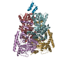
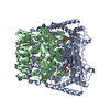
- Structure visualization
| Structure viewer | Molecule: MolmilJmol/JSmol |
|---|
- Downloads & links
-Download
| PDBx/mmCIF format | 3duf.cif.gz | 570.3 KB | Display | PDBx/mmCIF format |
|---|---|---|---|---|
| PDB format | pdb3duf.ent.gz | 454.9 KB | Display | PDB format |
| PDBx/mmJSON format | 3duf.json.gz | Tree view | PDBx/mmJSON format | |
| Others |  Other downloads Other downloads |
-Validation report
| Arichive directory | https://data.pdbj.org/pub/pdb/validation_reports/du/3dufftp://data.pdbj.org/pub/pdb/validation_reports/du/3duf | HTTPS FTP |
|---|
-Related structure data
| Related structure data |  3dv0C  3dvaC  1w85S S: Starting model for refinement C: citing same article ( |
|---|---|
| Similar structure data |


-Links
 PDBj
PDBj


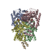
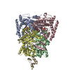
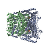
- Assembly
| Deposited unit | 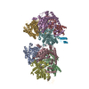
| |||||||||||||||||||||||||||||||||||||||||||||||||||||||||||||||||||||||||||||||||||||||||||||||||||||||||||||||||||||||||||||
|---|---|---|---|---|---|---|---|---|---|---|---|---|---|---|---|---|---|---|---|---|---|---|---|---|---|---|---|---|---|---|---|---|---|---|---|---|---|---|---|---|---|---|---|---|---|---|---|---|---|---|---|---|---|---|---|---|---|---|---|---|---|---|---|---|---|---|---|---|---|---|---|---|---|---|---|---|---|---|---|---|---|---|---|---|---|---|---|---|---|---|---|---|---|---|---|---|---|---|---|---|---|---|---|---|---|---|---|---|---|---|---|---|---|---|---|---|---|---|---|---|---|---|---|---|---|---|
| 1 | 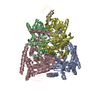
| |||||||||||||||||||||||||||||||||||||||||||||||||||||||||||||||||||||||||||||||||||||||||||||||||||||||||||||||||||||||||||||
| 2 | 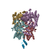
| |||||||||||||||||||||||||||||||||||||||||||||||||||||||||||||||||||||||||||||||||||||||||||||||||||||||||||||||||||||||||||||
| Unit cell |
| |||||||||||||||||||||||||||||||||||||||||||||||||||||||||||||||||||||||||||||||||||||||||||||||||||||||||||||||||||||||||||||
| Noncrystallographic symmetry (NCS) | NCS domain:
NCS domain segments: Component-ID: 1 / Refine code: 6
NCS ensembles :
|
-Components
-Pyruvate dehydrogenase E1 component subunit ... , 2 types, 8 molecules ACEGBDFH
| #1: Protein | Mass: 41518.176 Da / Num. of mol.: 4 Source method: isolated from a genetically manipulated source Source: (gene. exp.) Bacillus stearothermophilus (bacteria) / Gene: pdhA / Plasmid: pKBstE1a / Production host: References: UniProt: P21873, pyruvate dehydrogenase (acetyl-transferring) #2: Protein | Mass: 35495.633 Da / Num. of mol.: 4 Source method: isolated from a genetically manipulated source Source: (gene. exp.) Bacillus stearothermophilus (bacteria) / Gene: pdhB / Plasmid: pKBstE1b / Production host: References: UniProt: P21874, pyruvate dehydrogenase (acetyl-transferring) |
|---|
-Protein , 1 types, 2 molecules IJ
| #3: Protein | Mass: 46392.215 Da / Num. of mol.: 2 Source method: isolated from a genetically manipulated source Source: (gene. exp.) Bacillus stearothermophilus (bacteria) / Gene: pdhC / Plasmid: pKBstE1a / Production host: References: UniProt: P11961, dihydrolipoyllysine-residue acetyltransferase |
|---|
-Non-polymers , 4 types, 418 molecules 
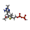


| #4: Chemical | ChemComp-MG /  Mass: 24.305 Da / Num. of mol.: 5 / Source method: obtained synthetically / Formula: Mg Mass: 24.305 Da / Num. of mol.: 5 / Source method: obtained synthetically / Formula: Mg#5: Chemical | ChemComp-R1T /  Mass: 467.371 Da / Num. of mol.: 4 / Source method: obtained synthetically / Formula: C15H23N3O8P2S Mass: 467.371 Da / Num. of mol.: 4 / Source method: obtained synthetically / Formula: C15H23N3O8P2S#6: Chemical |  Mass: 39.098 Da / Num. of mol.: 2 / Source method: obtained synthetically / Formula: K Mass: 39.098 Da / Num. of mol.: 2 / Source method: obtained synthetically / Formula: K#7: Water | ChemComp-HOH / | Mass: 18.015 Da / Num. of mol.: 407 / Source method: isolated from a natural source / Formula: H2O |
|---|
-Experimental details
-Experiment
| Experiment | Method: X-RAY DIFFRACTION / Number of used crystals: 1 |
|---|
- Sample preparation
| Crystal | Density Matthews: 1.86 Å3/Da / Density % sol: 33.72 % |
|---|---|
| Crystal grow | Temperature: 291.15 K / Method: vapor diffusion, sitting drop / pH: 5.5 Details: The protein solution was then mixed in 1:1 volume ratio of crystallization buffer consisting of 8-12 % mono-methyl ether polyethylene glycol (MME PEG) 5000, 0.1 M Na maleate pH 5.5, and the ...Details: The protein solution was then mixed in 1:1 volume ratio of crystallization buffer consisting of 8-12 % mono-methyl ether polyethylene glycol (MME PEG) 5000, 0.1 M Na maleate pH 5.5, and the droplet was left to equilibrate against a reservoir of neat crystallization buffer., VAPOR DIFFUSION, SITTING DROP, temperature 291.15K |
-Data collection
| Diffraction | Mean temperature: 100 K |
|---|---|
| Diffraction source | Source: SYNCHROTRON / Site: ESRF  / Beamline: ID23-1 / Wavelength: 0.9795 Å / Beamline: ID23-1 / Wavelength: 0.9795 Å |
| Detector | Type: MARMOSAIC 225 mm CCD / Detector: CCD / Date: Nov 11, 2005 / Details: mirror |
| Radiation | Monochromator: GRAPHITE / Protocol: SINGLE WAVELENGTH / Monochromatic (M) / Laue (L): M / Scattering type: x-ray |
| Radiation wavelength | Wavelength: 0.9795 Å / Relative weight: 1 |
| Reflection | Resolution: 2.5→66 Å / Num. all: 99540 / Num. obs: 99351 / % possible obs: 98.9 % / Observed criterion σ(F): 2.6 / Observed criterion σ(I): 2.6 / Redundancy: 3.7 % / Rmerge(I) obs: 0.111 / Rsym value: 0.111 / Net I/σ(I): 3.1 |
| Reflection shell | Resolution: 2.5→2.64 Å / Redundancy: 3.7 % / Rmerge(I) obs: 0.26 / Mean I/σ(I) obs: 5.4 / Rsym value: 0.302 / % possible all: 99.9 |
- Processing
| Software |
| ||||||||||||||||||||||||||||||||||||||||||||||||||||||||||||||||||||||||||||||||||||||||||||||||||||||||||||||||||||||||||||||||||||||||||||||||||||||||||||||||||||||||||
|---|---|---|---|---|---|---|---|---|---|---|---|---|---|---|---|---|---|---|---|---|---|---|---|---|---|---|---|---|---|---|---|---|---|---|---|---|---|---|---|---|---|---|---|---|---|---|---|---|---|---|---|---|---|---|---|---|---|---|---|---|---|---|---|---|---|---|---|---|---|---|---|---|---|---|---|---|---|---|---|---|---|---|---|---|---|---|---|---|---|---|---|---|---|---|---|---|---|---|---|---|---|---|---|---|---|---|---|---|---|---|---|---|---|---|---|---|---|---|---|---|---|---|---|---|---|---|---|---|---|---|---|---|---|---|---|---|---|---|---|---|---|---|---|---|---|---|---|---|---|---|---|---|---|---|---|---|---|---|---|---|---|---|---|---|---|---|---|---|---|---|---|
| Refinement | Method to determine structure: MOLECULAR REPLACEMENT Starting model: PDB entry 1W85 Resolution: 2.5→59.44 Å / Cor.coef. Fo:Fc: 0.941 / Cor.coef. Fo:Fc free: 0.888 / Isotropic thermal model: Isotropic / Cross valid method: THROUGHOUT / ESU R: 1.087 / ESU R Free: 0.328 / Stereochemistry target values: MAXIMUM LIKELIHOOD / Details: HYDROGENS HAVE BEEN ADDED IN THE RIDING POSITIONS
| ||||||||||||||||||||||||||||||||||||||||||||||||||||||||||||||||||||||||||||||||||||||||||||||||||||||||||||||||||||||||||||||||||||||||||||||||||||||||||||||||||||||||||
| Solvent computation | Ion probe radii: 0.8 Å / Shrinkage radii: 0.8 Å / VDW probe radii: 1.2 Å / Solvent model: MASK | ||||||||||||||||||||||||||||||||||||||||||||||||||||||||||||||||||||||||||||||||||||||||||||||||||||||||||||||||||||||||||||||||||||||||||||||||||||||||||||||||||||||||||
| Displacement parameters | Biso mean: 42.879 Å2
| ||||||||||||||||||||||||||||||||||||||||||||||||||||||||||||||||||||||||||||||||||||||||||||||||||||||||||||||||||||||||||||||||||||||||||||||||||||||||||||||||||||||||||
| Refine analyze |
| ||||||||||||||||||||||||||||||||||||||||||||||||||||||||||||||||||||||||||||||||||||||||||||||||||||||||||||||||||||||||||||||||||||||||||||||||||||||||||||||||||||||||||
| Refinement step | Cycle: LAST / Resolution: 2.5→59.44 Å
| ||||||||||||||||||||||||||||||||||||||||||||||||||||||||||||||||||||||||||||||||||||||||||||||||||||||||||||||||||||||||||||||||||||||||||||||||||||||||||||||||||||||||||
| Refine LS restraints |
| ||||||||||||||||||||||||||||||||||||||||||||||||||||||||||||||||||||||||||||||||||||||||||||||||||||||||||||||||||||||||||||||||||||||||||||||||||||||||||||||||||||||||||
| Refine LS restraints NCS | Refine-ID: X-RAY DIFFRACTION
| ||||||||||||||||||||||||||||||||||||||||||||||||||||||||||||||||||||||||||||||||||||||||||||||||||||||||||||||||||||||||||||||||||||||||||||||||||||||||||||||||||||||||||
| LS refinement shell | Resolution: 2.5→2.565 Å / Total num. of bins used: 20
|
