 Movie
Movie Controller

[English] 日本語
 Yorodumi
- PDB-3ex8: Complex structure of bacillus subtilis RibG reduction mechanism i... -
+ Open data

- Basic information
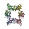
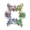
| Entry | Database: PDB / ID: 3ex8 | ||||||
|---|---|---|---|---|---|---|---|
| Title | Complex structure of bacillus subtilis RibG reduction mechanism in riboflavin biosynthesis | ||||||
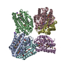
 Components Components | Riboflavin biosynthesis protein ribD | ||||||
 Keywords Keywords | HYDROLASE / OXIDOREDUCTASE / alpha/beta/alpha / deaminase domain / reductase domain / Metal-binding / Multifunctional enzyme / NADP / Riboflavin biosynthesis / Zinc | ||||||
| Function / homology |  Function and homology information Function and homology information5-amino-6-(5-phosphoribosylamino)uracil reductase / diaminohydroxyphosphoribosylaminopyrimidine deaminase / diaminohydroxyphosphoribosylaminopyrimidine deaminase activity / 5-amino-6-(5-phosphoribosylamino)uracil reductase activity / riboflavin biosynthetic process / NADP binding / zinc ion binding Similarity search - Function | ||||||
| Biological species |  | ||||||
| Method |  X-RAY DIFFRACTION / SYNCHROTRON / MOLECULAR REPLACEMENT / Resolution: 2.56 Å X-RAY DIFFRACTION / SYNCHROTRON / MOLECULAR REPLACEMENT / Resolution: 2.56 Å | ||||||
 Authors Authors | Chen, S.C. / Lin, Y.H. / Yu, H.C. / Liaw, S.H. | ||||||
 Citation Citation | Journal: J.Biol.Chem. / Year: 2009 Title: Complex structure of Bacillus subtilis RibG: the reduction mechanism during riboflavin biosynthesis. Authors: Chen, S.C. / Lin, Y.H. / Yu, H.C. / Liaw, S.H. #1: Journal: J.Biol.Chem. / Year: 2006 Title: Crystal structure of a bifunctional deaminase and reductase from Bacillus subtilis involved in riboflavin biosynthesis. Authors: Chen, S.C. / Chang, Y.C. / Lin, C.H. / Liaw, S.H. | ||||||
| History |
|
- Structure visualization
| Structure viewer | Molecule: MolmilJmol/JSmol |
|---|
- Downloads & links
-Download
| PDBx/mmCIF format | 3ex8.cif.gz | 281.4 KB | Display | PDBx/mmCIF format |
|---|---|---|---|---|
| PDB format | pdb3ex8.ent.gz | 226.7 KB | Display | PDB format |
| PDBx/mmJSON format | 3ex8.json.gz | Tree view | PDBx/mmJSON format | |
| Others |  Other downloads Other downloads |
-Validation report
| Arichive directory | https://data.pdbj.org/pub/pdb/validation_reports/ex/3ex8ftp://data.pdbj.org/pub/pdb/validation_reports/ex/3ex8 | HTTPS FTP |
|---|
-Related structure data
| Related structure data | 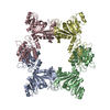 2b3zS S: Starting model for refinement |
|---|---|
| Similar structure data |


-Links
 PDBj
PDBj
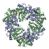
- Assembly
| Deposited unit | 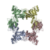
| ||||||||
|---|---|---|---|---|---|---|---|---|---|
| 1 |
| ||||||||
| Unit cell |
|
-Components
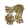
| #1: Protein | Mass: 40759.758 Da / Num. of mol.: 4 Source method: isolated from a genetically manipulated source Source: (gene. exp.) References: UniProt: P17618, diaminohydroxyphosphoribosylaminopyrimidine deaminase, 5-amino-6-(5-phosphoribosylamino)uracil reductase #2: Chemical | ChemComp-ZN /   Mass: 65.409 Da / Num. of mol.: 4 / Source method: obtained synthetically / Formula: Zn Mass: 65.409 Da / Num. of mol.: 4 / Source method: obtained synthetically / Formula: Zn#3: Chemical | ChemComp-AIF / [( | 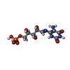  Mass: 354.211 Da / Num. of mol.: 1 / Source method: obtained synthetically / Formula: C9H15N4O9P Mass: 354.211 Da / Num. of mol.: 1 / Source method: obtained synthetically / Formula: C9H15N4O9P#4: Water | ChemComp-HOH / |  Mass: 18.015 Da / Num. of mol.: 191 / Source method: isolated from a natural source / Formula: H2O Mass: 18.015 Da / Num. of mol.: 191 / Source method: isolated from a natural source / Formula: H2O |
|---|
-Experimental details
-Experiment
| Experiment | Method: X-RAY DIFFRACTION / Number of used crystals: 1 |
|---|
- Sample preparation
| Crystal | Density Matthews: 2.71 Å3/Da / Density % sol: 54.64 % |
|---|---|
| Crystal grow | Temperature: 295 K / Method: vapor diffusion, hanging drop / pH: 7.5 Details: 28 % PEG 400, 200mM CaCl2, 100mM HEPES pH 7.5, VAPOR DIFFUSION, HANGING DROP, temperature 295K |
-Data collection
| Diffraction | Mean temperature: 100 K |
|---|---|
| Diffraction source | Source: SYNCHROTRON / Site: NSRRC  / Beamline: BL13B1 / Wavelength: 1 Å / Beamline: BL13B1 / Wavelength: 1 Å |
| Detector | Type: ADSC QUANTUM 315 / Detector: CCD / Date: Nov 6, 2005 |
| Radiation | Protocol: SINGLE WAVELENGTH / Monochromatic (M) / Laue (L): M / Scattering type: x-ray |
| Radiation wavelength | Wavelength: 1 Å / Relative weight: 1 |
| Reflection | Resolution: 2.56→50 Å / Num. all: 57889 / Num. obs: 56499 / % possible obs: 97.7 % / Observed criterion σ(F): 0 / Observed criterion σ(I): 0 / Redundancy: 8.3 % / Rmerge(I) obs: 0.062 / Net I/σ(I): 26.2 |
| Reflection shell | Resolution: 2.56→2.65 Å / Redundancy: 4.1 % / Rmerge(I) obs: 0.426 / Mean I/σ(I) obs: 2.7 / Num. unique all: 5437 / % possible all: 95.4 |
- Processing
| Software |
| |||||||||||||||||||||||||
|---|---|---|---|---|---|---|---|---|---|---|---|---|---|---|---|---|---|---|---|---|---|---|---|---|---|---|
| Refinement | Method to determine structure: MOLECULAR REPLACEMENT Starting model: 2B3Z Resolution: 2.56→50 Å / Isotropic thermal model: Anisotropic / Cross valid method: THROUGHOUT / σ(F): 0 / σ(I): 0 / Stereochemistry target values: Engh & Huber
| |||||||||||||||||||||||||
| Displacement parameters |
| |||||||||||||||||||||||||
| Refine analyze |
| |||||||||||||||||||||||||
| Refinement step | Cycle: LAST / Resolution: 2.56→50 Å
| |||||||||||||||||||||||||
| Refine LS restraints |
| |||||||||||||||||||||||||
| LS refinement shell | Resolution: 2.56→2.65 Å
|
