 Movie
Movie Controller

[English] 日本語
 Yorodumi
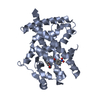
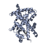
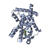
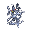
- PDB-3et0: Structure of PPARgamma with 3-(5-Methoxy-1H-indol-3-yl)-propionic acid -
+ Open data

- Basic information
| Entry | Database: PDB / ID: 3et0 | ||||||
|---|---|---|---|---|---|---|---|
| Title | Structure of PPARgamma with 3-(5-Methoxy-1H-indol-3-yl)-propionic acid | ||||||
 Components Components | Peroxisome proliferator-activated receptor gamma | ||||||
 Keywords Keywords | TRANSCRIPTION / PPAR / PPARg / PPARgamma / Drug Discovery / Diabetes / adiponectin / metabolic disease / fragment-based drug discovery / scaffold-based drug discovery / Activator / Alternative splicing / Diabetes mellitus / Disease mutation / DNA-binding / Metal-binding / Nucleus / Obesity / Phosphoprotein / Polymorphism / Receptor / Transcription regulation / Zinc / Zinc-finger | ||||||
| Function / homology |  Function and homology information Function and homology informationprostaglandin receptor activity / : / negative regulation of receptor signaling pathway via STAT / MECP2 regulates transcription factors / negative regulation of vascular endothelial cell proliferation / negative regulation of extracellular matrix assembly / negative regulation of connective tissue replacement involved in inflammatory response wound healing / positive regulation of cholesterol transport / negative regulation of cellular response to transforming growth factor beta stimulus / arachidonate binding ...prostaglandin receptor activity / : / negative regulation of receptor signaling pathway via STAT / MECP2 regulates transcription factors / negative regulation of vascular endothelial cell proliferation / negative regulation of extracellular matrix assembly / negative regulation of connective tissue replacement involved in inflammatory response wound healing / positive regulation of cholesterol transport / negative regulation of cellular response to transforming growth factor beta stimulus / arachidonate binding / positive regulation of adiponectin secretion / negative regulation of cardiac muscle hypertrophy in response to stress / DNA binding domain binding / lipoprotein transport / positive regulation of vascular associated smooth muscle cell apoptotic process / WW domain binding / positive regulation of fatty acid metabolic process / STAT family protein binding / response to lipid / negative regulation of type II interferon-mediated signaling pathway / LBD domain binding / negative regulation of cholesterol storage / negative regulation of SMAD protein signal transduction / lipid homeostasis / E-box binding / alpha-actinin binding / R-SMAD binding / negative regulation of vascular associated smooth muscle cell proliferation / monocyte differentiation / negative regulation of blood vessel endothelial cell migration / white fat cell differentiation / negative regulation of macrophage derived foam cell differentiation / cellular response to low-density lipoprotein particle stimulus / negative regulation of lipid storage / negative regulation of BMP signaling pathway / positive regulation of cholesterol efflux / cell fate commitment / negative regulation of osteoblast differentiation / negative regulation of mitochondrial fission / positive regulation of fat cell differentiation / BMP signaling pathway / long-chain fatty acid transport / nuclear retinoid X receptor binding / Transcriptional regulation of brown and beige adipocyte differentiation by EBF2 / retinoic acid receptor signaling pathway / cell maturation / negative regulation of MAPK cascade / intracellular receptor signaling pathway / hormone-mediated signaling pathway / positive regulation of adipose tissue development / peroxisome proliferator activated receptor signaling pathway / response to nutrient / epithelial cell differentiation / regulation of cellular response to insulin stimulus / peptide binding / negative regulation of miRNA transcription / negative regulation of angiogenesis / placenta development / Regulation of PTEN gene transcription / positive regulation of apoptotic signaling pathway / transcription coregulator binding / SUMOylation of intracellular receptors / negative regulation of smooth muscle cell proliferation / mRNA transcription by RNA polymerase II / negative regulation of transforming growth factor beta receptor signaling pathway / fatty acid metabolic process / PPARA activates gene expression / regulation of circadian rhythm / Nuclear Receptor transcription pathway / Transcriptional regulation of white adipocyte differentiation / lipid metabolic process / positive regulation of miRNA transcription / regulation of blood pressure / negative regulation of inflammatory response / DNA-binding transcription repressor activity, RNA polymerase II-specific / RNA polymerase II transcription regulator complex / nuclear receptor activity / cellular response to insulin stimulus / rhythmic process / glucose homeostasis / MLL4 and MLL3 complexes regulate expression of PPARG target genes in adipogenesis and hepatic steatosis / double-stranded DNA binding / DNA-binding transcription activator activity, RNA polymerase II-specific / cellular response to hypoxia / DNA-binding transcription factor binding / sequence-specific DNA binding / nucleic acid binding / DNA-binding transcription factor activity, RNA polymerase II-specific / cell differentiation / receptor complex / transcription cis-regulatory region binding / RNA polymerase II cis-regulatory region sequence-specific DNA binding / DNA-binding transcription factor activity / negative regulation of gene expression / innate immune response / negative regulation of DNA-templated transcription / intracellular membrane-bounded organelle / chromatin binding / positive regulation of gene expression / regulation of transcription by RNA polymerase II Similarity search - Function | ||||||
| Biological species |  Homo sapiens (human) Homo sapiens (human) | ||||||
| Method |  X-RAY DIFFRACTION / SYNCHROTRON / MOLECULAR REPLACEMENT / Resolution: 2.4 Å X-RAY DIFFRACTION / SYNCHROTRON / MOLECULAR REPLACEMENT / Resolution: 2.4 Å | ||||||
 Authors Authors | Zhang, K.Y.J. / Wang, W. | ||||||
 Citation Citation | Journal: Proc.Natl.Acad.Sci.USA / Year: 2009 Title: Scaffold-based discovery of indeglitazar, a PPAR pan-active anti-diabetic agent Authors: Artis, D.R. / Lin, J.J. / Zhang, C. / Wang, W. / Mehra, U. / Perreault, M. / Erbe, D. / Krupka, H.I. / England, B.P. / Arnold, J. / Plotnikov, A.N. / Marimuthu, A. / Nguyen, H. / Will, S. / ...Authors: Artis, D.R. / Lin, J.J. / Zhang, C. / Wang, W. / Mehra, U. / Perreault, M. / Erbe, D. / Krupka, H.I. / England, B.P. / Arnold, J. / Plotnikov, A.N. / Marimuthu, A. / Nguyen, H. / Will, S. / Signaevsky, M. / Kral, J. / Cantwell, J. / Settachatgull, C. / Yan, D.S. / Fong, D. / Oh, A. / Shi, S. / Womack, P. / Powell, B. / Habets, G. / West, B.L. / Zhang, K.Y. / Milburn, M.V. / Vlasuk, G.P. / Hirth, K.P. / Nolop, K. / Bollag, G. / Ibrahim, P.N. / Tobin, J.F. | ||||||
| History |
|
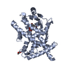
- Structure visualization
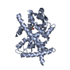
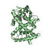
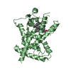
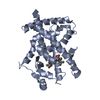
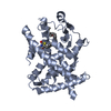
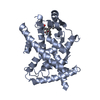
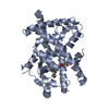
| Structure viewer | Molecule: MolmilJmol/JSmol |
|---|
- Downloads & links
-Download
| PDBx/mmCIF format | 3et0.cif.gz | 119.1 KB | Display | PDBx/mmCIF format |
|---|---|---|---|---|
| PDB format | pdb3et0.ent.gz | 92.4 KB | Display | PDB format |
| PDBx/mmJSON format | 3et0.json.gz | Tree view | PDBx/mmJSON format | |
| Others |  Other downloads Other downloads |
-Validation report
| Summary document | 3et0_validation.pdf.gz | 480 KB | Display | wwPDB validaton report |
|---|---|---|---|---|
| Full document | 3et0_full_validation.pdf.gz | 493.6 KB | Display | |
| Data in XML | 3et0_validation.xml.gz | 23.1 KB | Display | |
| Data in CIF | 3et0_validation.cif.gz | 30.9 KB | Display | |
| Arichive directory | https://data.pdbj.org/pub/pdb/validation_reports/et/3et0ftp://data.pdbj.org/pub/pdb/validation_reports/et/3et0 | HTTPS FTP |
-Related structure data
| Related structure data |  3et1C  3et2C  3et3C 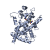 2prgS C: citing same article ( S: Starting model for refinement |
|---|---|
| Similar structure data |


-Links
 PDBj
PDBj

- Assembly
| Deposited unit | 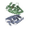
| ||||||||
|---|---|---|---|---|---|---|---|---|---|
| 1 | 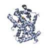
| ||||||||
| 2 | 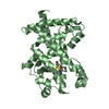
| ||||||||
| Unit cell |
|
-Components
| #1: Protein | Mass: 33375.688 Da / Num. of mol.: 2 / Fragment: LIGAND BINDING DOMAIN Source method: isolated from a genetically manipulated source Source: (gene. exp.) Homo sapiens (human) / Gene: PPARG, NR1C3 / Plasmid: pET-28 / Production host:  #2: Chemical | ChemComp-ET0 / |   Mass: 219.237 Da / Num. of mol.: 1 / Source method: obtained synthetically / Formula: C12H13NO3 Mass: 219.237 Da / Num. of mol.: 1 / Source method: obtained synthetically / Formula: C12H13NO3#3: Sugar | ChemComp-GLC / |   Type: D-saccharide, alpha linking / Mass: 180.156 Da / Num. of mol.: 1 Type: D-saccharide, alpha linking / Mass: 180.156 Da / Num. of mol.: 1Source method: isolated from a genetically manipulated source Formula: C6H12O6 #4: Water | ChemComp-HOH / |  Mass: 18.015 Da / Num. of mol.: 98 / Source method: isolated from a natural source / Formula: H2O Mass: 18.015 Da / Num. of mol.: 98 / Source method: isolated from a natural source / Formula: H2OHas protein modification | Y | |
|---|
-Experimental details
-Experiment
| Experiment | Method: X-RAY DIFFRACTION / Number of used crystals: 1 |
|---|
- Sample preparation
| Crystal | Density Matthews: 2.55 Å3/Da / Density % sol: 51.75 % |
|---|---|
| Crystal grow | Temperature: 277 K / Method: vapor diffusion, sitting drop / pH: 7.5 Details: The purified PPARg ligand binding domain (LBD) protein was diluated to 12 mg/ml and 1mM of compound 1 was added to the protein prior to crystallization by mixing equal volumes of ...Details: The purified PPARg ligand binding domain (LBD) protein was diluated to 12 mg/ml and 1mM of compound 1 was added to the protein prior to crystallization by mixing equal volumes of protein/compound sample with reservoir solution containing 0.9 M sodium citrate and 0.1 M 4-(2-hydroxyethyl)-1-piperazineethanesulfonic acid (HEPES) at pH 7.5, VAPOR DIFFUSION, SITTING DROP, temperature 277K |
-Data collection
| Diffraction | Mean temperature: 180 K |
|---|---|
| Diffraction source | Source: SYNCHROTRON / Site: ALS  / Beamline: 5.0.1 / Wavelength: 1.1 Å / Beamline: 5.0.1 / Wavelength: 1.1 Å |
| Detector | Type: ADSC QUANTUM 210 / Detector: CCD / Date: Jun 5, 2002 |
| Radiation | Protocol: SINGLE WAVELENGTH / Monochromatic (M) / Laue (L): M / Scattering type: x-ray |
| Radiation wavelength | Wavelength: 1.1 Å / Relative weight: 1 |
| Reflection | Resolution: 2.4→50 Å / Num. all: 27794 / Num. obs: 26349 / % possible obs: 0.948 % / Observed criterion σ(F): 0 / Observed criterion σ(I): 0 / Redundancy: 2.6 % / Rsym value: 0.046 |
| Reflection shell | Resolution: 2.4→2.55 Å / Redundancy: 2.5 % / Num. unique all: 3206 / Rsym value: 0.391 / % possible all: 94.2 |
- Processing
| Software |
| |||||||||||||||||||||||||||||||||||||||||||||||||||||||||||||||||||||||||||
|---|---|---|---|---|---|---|---|---|---|---|---|---|---|---|---|---|---|---|---|---|---|---|---|---|---|---|---|---|---|---|---|---|---|---|---|---|---|---|---|---|---|---|---|---|---|---|---|---|---|---|---|---|---|---|---|---|---|---|---|---|---|---|---|---|---|---|---|---|---|---|---|---|---|---|---|---|
| Refinement | Method to determine structure: MOLECULAR REPLACEMENT Starting model: PDB entry 2PRG Resolution: 2.4→29.188 Å / SU ML: 0.4 / σ(F): 1.34 / σ(I): 0 / Stereochemistry target values: ML
| |||||||||||||||||||||||||||||||||||||||||||||||||||||||||||||||||||||||||||
| Solvent computation | Shrinkage radii: 0.9 Å / VDW probe radii: 1.11 Å / Solvent model: FLAT BULK SOLVENT MODEL / Bsol: 70.362 Å2 / ksol: 0.333 e/Å3 | |||||||||||||||||||||||||||||||||||||||||||||||||||||||||||||||||||||||||||
| Refinement step | Cycle: LAST / Resolution: 2.4→29.188 Å
| |||||||||||||||||||||||||||||||||||||||||||||||||||||||||||||||||||||||||||
| LS refinement shell |
| |||||||||||||||||||||||||||||||||||||||||||||||||||||||||||||||||||||||||||
| Refinement TLS params. | Method: refined / Refine-ID: X-RAY DIFFRACTION
| |||||||||||||||||||||||||||||||||||||||||||||||||||||||||||||||||||||||||||
| Refinement TLS group |
|
