 Movie
Movie Controller

[English] 日本語
 Yorodumi
- PDB-3esi: Crystal structure of an uncharacterized protein from Erwinia caro... -
+ Open data

- Basic information
| Entry | Database: PDB / ID: 3esi | ||||||
|---|---|---|---|---|---|---|---|
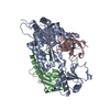
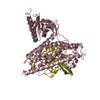
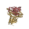
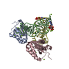
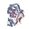
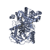
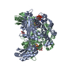
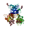
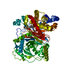
| Title | Crystal structure of an uncharacterized protein from Erwinia carotovora subsp. atroseptica. Northeast Structural Genomics target EwR179 | ||||||
 Components Components | uncharacterized protein | ||||||
 Keywords Keywords | structural genomics / unknown function / protein from Erwinia carotovora subsp. atroseptica (Pectobacterium atrosepticum) / PSI-2 / Protein Structure Initiative / Northeast Structural Genomics Consortium / NESG | ||||||
| Function / homology | : / Dehydrase, ECs4332, predicted / ApeI dehydratase / Hotdog Thioesterase / Thiol Ester Dehydrase; Chain A / HotDog domain superfamily / Roll / Alpha Beta / ApeI dehydratase-like domain-containing protein Function and homology information Function and homology information | ||||||
| Biological species |  Pectobacterium atrosepticum (bacteria) Pectobacterium atrosepticum (bacteria) | ||||||
| Method |  X-RAY DIFFRACTION / SYNCHROTRON / SAD / Resolution: 2.5 Å X-RAY DIFFRACTION / SYNCHROTRON / SAD / Resolution: 2.5 Å | ||||||
 Authors Authors | Seetharaman, J. / Lew, S. / Wang, H. / Janjua, H. / Foote, E.L. / Xiao, R. / Nair, R. / Everett, J.K. / Acton, T.B. / Rost, B. ...Seetharaman, J. / Lew, S. / Wang, H. / Janjua, H. / Foote, E.L. / Xiao, R. / Nair, R. / Everett, J.K. / Acton, T.B. / Rost, B. / Montelione, G.T. / Hunt, J.F. / Tong, L. / Northeast Structural Genomics Consortium (NESG) | ||||||
 Citation Citation | Journal: To be Published Title: Crystal structure of an uncharacterized protein from Erwinia carotovora subsp. atroseptica. Northeast Structural Genomics target EwR179 Authors: Seetharaman, J. / Lew, S. / Wang, H. / Janjua, H. / Foote, E.L. / Xiao, R. / Nair, R. / Everett, J.K. / Acton, T.B. / Rost, B. / Montelione, G.T. / Hunt, J.F. / Tong, L. | ||||||
| History |
|
- Structure visualization
| Structure viewer | Molecule: MolmilJmol/JSmol |
|---|
- Downloads & links
-Download
| PDBx/mmCIF format | 3esi.cif.gz | 106.7 KB | Display | PDBx/mmCIF format |
|---|---|---|---|---|
| PDB format | pdb3esi.ent.gz | 83.9 KB | Display | PDB format |
| PDBx/mmJSON format | 3esi.json.gz | Tree view | PDBx/mmJSON format | |
| Others |  Other downloads Other downloads |
-Validation report
| Arichive directory | https://data.pdbj.org/pub/pdb/validation_reports/es/3esiftp://data.pdbj.org/pub/pdb/validation_reports/es/3esi | HTTPS FTP |
|---|
-Related structure data
| Similar structure data | |
|---|---|
| Other databases |

-Links
 PDBj
PDBj
- Assembly
| Deposited unit | 
| ||||||||
|---|---|---|---|---|---|---|---|---|---|
| 1 |
| ||||||||
| Unit cell |
|
-Components
| #1: Protein | Mass: 14651.850 Da / Num. of mol.: 4 Source method: isolated from a genetically manipulated source Source: (gene. exp.) Pectobacterium atrosepticum (bacteria) / Gene: ECA4500 / Plasmid: PET 21 / Production host: #2: Water | ChemComp-HOH / |  Mass: 18.015 Da / Num. of mol.: 74 / Source method: isolated from a natural source / Formula: H2O Mass: 18.015 Da / Num. of mol.: 74 / Source method: isolated from a natural source / Formula: H2O |
|---|
-Experimental details
-Experiment
| Experiment | Method: X-RAY DIFFRACTION / Number of used crystals: 1 |
|---|
- Sample preparation
| Crystal | Density Matthews: 3.16 Å3/Da / Density % sol: 61.14 % |
|---|---|
| Crystal grow | Temperature: 293 K / pH: 5.6 Details: peg4k, 200mM NH4Acetate, 100mM Na3citrate ph5.6, temperature 293K |
-Data collection
| Diffraction |
| |||||||||||||||
|---|---|---|---|---|---|---|---|---|---|---|---|---|---|---|---|---|
| Diffraction source |
| |||||||||||||||
| Detector | Type: ADSC QUANTUM 210 / Detector: CCD / Date: Jul 24, 2008 / Details: Mirrors | |||||||||||||||
| Radiation | Protocol: SINGLE WAVELENGTH / Monochromatic (M) / Laue (L): M / Scattering type: x-ray | |||||||||||||||
| Radiation wavelength | Wavelength: 0.979 Å / Relative weight: 1 | |||||||||||||||
| Reflection | Resolution: 2.5→50 Å / Num. obs: 49294 / % possible obs: 100 % / Observed criterion σ(F): 0 / Observed criterion σ(I): 0 / Redundancy: 6.5 % / Biso Wilson estimate: 44.1 Å2 / Rmerge(I) obs: 0.054 / Rsym value: 0.046 / Net I/σ(I): 14 | |||||||||||||||
| Reflection shell | Resolution: 2.5→2.59 Å / Redundancy: 6.5 % / Rmerge(I) obs: 0.249 / Mean I/σ(I) obs: 14 / Num. unique all: 4932 / Rsym value: 0.196 / % possible all: 100 |

- Processing
| Software |
| ||||||||||||||||||||
|---|---|---|---|---|---|---|---|---|---|---|---|---|---|---|---|---|---|---|---|---|---|
| Refinement | Method to determine structure: SAD / Resolution: 2.5→35.05 Å / Rfactor Rfree error: 0.006 / Data cutoff high absF: 171847.19 / Data cutoff low absF: 0 / Isotropic thermal model: RESTRAINED / Cross valid method: THROUGHOUT / σ(F): 0 / Stereochemistry target values: Engh & Huber / Details: BULK SOLVENT MODEL USED
| ||||||||||||||||||||
| Solvent computation | Solvent model: FLAT MODEL / Bsol: 49.9999 Å2 / ksol: 0.4 e/Å3 | ||||||||||||||||||||
| Displacement parameters | Biso mean: 46.4 Å2
| ||||||||||||||||||||
| Refine analyze |
| ||||||||||||||||||||
| Refinement step | Cycle: LAST / Resolution: 2.5→35.05 Å
| ||||||||||||||||||||
| Refine LS restraints |
| ||||||||||||||||||||
| LS refinement shell | Resolution: 2.5→2.66 Å / Rfactor Rfree error: 0.018 / Total num. of bins used: 6
| ||||||||||||||||||||
| Xplor file |
|
