 Movie
Movie Controller

[English] 日本語
 Yorodumi
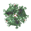
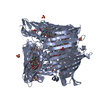
- PDB-3eoj: Fmo protein from Prosthecochloris Aestuarii 2K AT 1.3A Resolution -
+ Open data

- Basic information
| Entry | Database: PDB / ID: 3eoj | ||||||
|---|---|---|---|---|---|---|---|
| Title | Fmo protein from Prosthecochloris Aestuarii 2K AT 1.3A Resolution | ||||||
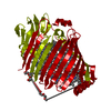
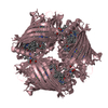
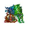
 Components Components | Bacteriochlorophyll a protein | ||||||
 Keywords Keywords | PHOTOSYNTHESIS / EXCITATION ENERGY TRANSFER / BETA SHEET / GAMMA-TURNS / Bacteriochlorophyll / Chlorophyll / Chromophore / Electron transport / Magnesium / Metal-binding / Reaction center / Transport | ||||||
| Function / homology |  Function and homology information Function and homology information | ||||||
| Biological species |  Prosthecochloris aestuarii 2K (bacteria) Prosthecochloris aestuarii 2K (bacteria) | ||||||
| Method |  X-RAY DIFFRACTION / SYNCHROTRON / MOLECULAR REPLACEMENT / Resolution: 1.3 Å X-RAY DIFFRACTION / SYNCHROTRON / MOLECULAR REPLACEMENT / Resolution: 1.3 Å | ||||||
 Authors Authors | Tronrud, D.E. / Wen, J. / Gay, L. / Blankenship, R.E. | ||||||
 Citation Citation | Journal: Photosynth.Res. / Year: 2009 Title: The structural basis for the difference in absorbance spectra for the FMO antenna protein from various green sulfur bacteria. Authors: Tronrud, D.E. / Wen, J. / Gay, L. / Blankenship, R.E. #1: Journal: The Photosynthetic Reaction Center / Year: 1993Title: Refinement of the Structure of a Water-Soluble Antenna Complex from Green Photosynthetic Bacteria by Incorporation of the Chemically Determined Amino Acid Sequence Authors: Tronrud, D.E. / Matthews, B.W. #2: Journal: J.Mol.Biol. / Year: 1986 Title: Structure and X-Ray Amino Acid Sequence of a Bacteriochlorophyll a Protein from Prosthecochloris Aestuarii Refined at 1.9 A Resolution Authors: Tronrud, D.E. / Schmid, M.F. / Matthews, B.W. #3: Journal: Chem.Scr. / Year: 1983Title: Structural Studies of a Bacteriochlorophyll-Containing Protein Authors: Schmid, M.F. / Tronrud, D.E. / Matthews, B.W. #4: Journal: Lipid-Protein Interactions / Year: 1982Title: Lipid-Protein Interactions in a Bacteriochlorophyll-Containing Protein Authors: Matthews, B.W. #5: Journal: Acc.Chem.Res. / Year: 1980Title: Structure of a Green Bacteriochlorophyll Protein Authors: Matthews, B.W. / Fenna, R.E. #6: Journal: J.Mol.Biol. / Year: 1979 Title: Structure of a Bacteriochlorophyll A-Protein from the Green Photosynthetic Bacterium Prosthecochloris Aestuarii Authors: Matthews, B.W. / Fenna, R.E. / Bolognesi, M.C. / Schmid, M.F. / Olson, J.M. #7: Journal: The Photosynthetic Bacteria / Year: 1978Title: Bacteriochlorophyll A-Protein from Green Bacteria Authors: Olson, J.M. #8: Journal: Biochem.Biophys.Res.Commun. / Year: 1977 Title: Atomic Coordinates for the Chlorophyll Core of a Bacteriochlorophyll A-Protein from Green Photosynthetic Bacteria Authors: Fenna, R.E. / Ten Eyck, L.F. / Matthews, B.W. #9: Journal: J.Ultrastruct.Res. / Year: 1977 Title: An Evaluation of Electron Micrographs of Bacteriochlorophyll A-Protein Crystals in Terms of the Structure Determined by X-Ray Crystallography Authors: Matthews, B.W. / Fenna, R.E. / Remington, S.J. #10: Journal: Brookhaven Symp.Biol. / Year: 1976Title: Structure of a Bacteriochlorophyll A-Protein from Prosthecochloris Aestuarii Authors: Fenna, R.E. / Matthews, B.W. #11: Journal: Nature / Year: 1975Title: Chlorophyll Arrangement in a Bacteriochlorophyll Protein from Chlorobium Limicola Authors: Fenna, R.E. / Matthews, B.W. | ||||||
| History |
|
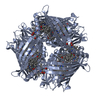
- Structure visualization
| Structure viewer | Molecule: MolmilJmol/JSmol |
|---|
- Downloads & links
-Download
| PDBx/mmCIF format | 3eoj.cif.gz | 217 KB | Display | PDBx/mmCIF format |
|---|---|---|---|---|
| PDB format | pdb3eoj.ent.gz | 176.1 KB | Display | PDB format |
| PDBx/mmJSON format | 3eoj.json.gz | Tree view | PDBx/mmJSON format | |
| Others |  Other downloads Other downloads |
-Validation report
| Arichive directory | https://data.pdbj.org/pub/pdb/validation_reports/eo/3eojftp://data.pdbj.org/pub/pdb/validation_reports/eo/3eoj | HTTPS FTP |
|---|
-Related structure data
| Related structure data |  3eniC  1bcl 2bcl S: Starting model for refinement C: citing same article ( |
|---|---|
| Similar structure data |

-Links
 PDBj
PDBj
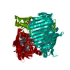
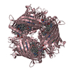
- Assembly
| Deposited unit | 
| ||||||||||||
|---|---|---|---|---|---|---|---|---|---|---|---|---|---|
| 1 | 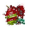
| ||||||||||||
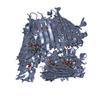
| Unit cell |
| ||||||||||||
| Components on special symmetry positions |
|
-Components
-Protein , 1 types, 1 molecules A
| #1: Protein | Mass: 40282.168 Da / Num. of mol.: 1 / Source method: isolated from a natural source / Details: ANTENNA COMPLEX / Source: (natural) Prosthecochloris aestuarii 2K (bacteria) / References: UniProt: P11741 |
|---|
-Non-polymers , 5 types, 431 molecules 


| #2: Chemical | ChemComp-BCL /  Mass: 911.504 Da / Num. of mol.: 8 / Source method: obtained synthetically / Formula: C55H74MgN4O6 Mass: 911.504 Da / Num. of mol.: 8 / Source method: obtained synthetically / Formula: C55H74MgN4O6#3: Chemical | ChemComp-EDO /  Mass: 62.068 Da / Num. of mol.: 15 / Source method: obtained synthetically / Formula: C2H6O2 Mass: 62.068 Da / Num. of mol.: 15 / Source method: obtained synthetically / Formula: C2H6O2#4: Chemical | ChemComp-NA / |  Mass: 22.990 Da / Num. of mol.: 1 / Source method: obtained synthetically / Formula: Na Mass: 22.990 Da / Num. of mol.: 1 / Source method: obtained synthetically / Formula: Na#5: Chemical | ChemComp-NH4 / |  Mass: 18.038 Da / Num. of mol.: 1 / Source method: obtained synthetically / Formula: H4N Mass: 18.038 Da / Num. of mol.: 1 / Source method: obtained synthetically / Formula: H4N#6: Water | ChemComp-HOH / | Mass: 18.015 Da / Num. of mol.: 406 / Source method: isolated from a natural source / Formula: H2O |
|---|
-Details
| Sequence details | ACCORDING TO THE AUTHORS THE DATABASE SEQUENCE IS IN ERROR |
|---|
-Experimental details
-Experiment
| Experiment | Method: X-RAY DIFFRACTION / Number of used crystals: 1 |
|---|
- Sample preparation
| Crystal | Density Matthews: 4.35 Å3/Da / Density % sol: 71.75 % |
|---|---|
| Crystal grow | Temperature: 298 K / Method: vapor diffusion, hanging drop / pH: 7.8 Details: Well Solution: 10mM Tris/HCL pH 8.0, 1.0M NaCl, 10% w/v (NH4)2SO4. Protein Solution: 10mM Tris/HCL pH 7.8 1.0M NaCl at 14mg/ml of complex. Drop composed of 5ul of protein solution and 3ul of ...Details: Well Solution: 10mM Tris/HCL pH 8.0, 1.0M NaCl, 10% w/v (NH4)2SO4. Protein Solution: 10mM Tris/HCL pH 7.8 1.0M NaCl at 14mg/ml of complex. Drop composed of 5ul of protein solution and 3ul of well solution., VAPOR DIFFUSION, HANGING DROP, temperature 298K |
-Data collection
| Diffraction | Mean temperature: 100 K | |||||||||
|---|---|---|---|---|---|---|---|---|---|---|
| Diffraction source | Source: SYNCHROTRON / Site: ALS  / Beamline: 8.2.2 / Wavelength: 1 / Wavelength: 1 Å / Beamline: 8.2.2 / Wavelength: 1 / Wavelength: 1 Å | |||||||||
| Detector | Type: ADSC QUANTUM 315 / Detector: CCD / Date: Jan 23, 2007 / Details: MIRRORS | |||||||||
| Radiation | Monochromator: DOUBLE CRYSTAL, SI(111) / Protocol: SINGLE WAVELENGTH / Monochromatic (M) / Laue (L): M / Scattering type: x-ray | |||||||||
| Radiation wavelength |
| |||||||||
| Reflection | Resolution: 1.3→50 Å / Num. all: 168292 / Num. obs: 168292 / % possible obs: 99.9 % / Observed criterion σ(F): 0 / Observed criterion σ(I): 0 / Redundancy: 7.8 % / Biso Wilson estimate: 15.6 Å2 / Rmerge(I) obs: 0.074 / Net I/σ(I): 26.5 | |||||||||
| Reflection shell | Resolution: 1.3→1.35 Å / Redundancy: 6.2 % / Rmerge(I) obs: 0.596 / Mean I/σ(I) obs: 2.77 / Num. unique all: 16812 / % possible all: 100 |
- Processing
| Software |
| |||||||||||||||||||||||||||||||||
|---|---|---|---|---|---|---|---|---|---|---|---|---|---|---|---|---|---|---|---|---|---|---|---|---|---|---|---|---|---|---|---|---|---|---|
| Refinement | Method to determine structure: MOLECULAR REPLACEMENT Starting model: PDB entry 1BCL 1bcl Resolution: 1.3→50 Å / Num. parameters: 37346 / Num. restraintsaints: 52424 / Isotropic thermal model: anisotropic / Cross valid method: FREE R / σ(F): 0 / σ(I): 0 / Stereochemistry target values: Engh & Huber Details: WEIGHTED FULL MATRIX LEAST SQUARES PROCEDURE. THE BOND LENGTHS AND ANGLES OF ALL BCL MOLECULES, EXCEPT FOR 378, WERE RESTRAINED TO THE GROUP AVERAGES CALCULATED FROM THESE SEVEN MOLECULES. ...Details: WEIGHTED FULL MATRIX LEAST SQUARES PROCEDURE. THE BOND LENGTHS AND ANGLES OF ALL BCL MOLECULES, EXCEPT FOR 378, WERE RESTRAINED TO THE GROUP AVERAGES CALCULATED FROM THESE SEVEN MOLECULES. NO EXTERNAL LIBRARY WAS USED. THE BOND LENGTHS AND ANGLES OF 378 WERE RESTRAINED TO THE AVERAGE VALUES OF THE OTHER SEVEN BCL MOLECULES
| |||||||||||||||||||||||||||||||||
| Solvent computation | Solvent model: MOEWS & KRETSINGER, J.MOL.BIOL.91(1973)201-228 Bsol: 3.31935 Å2 / ksol: 0.8587 e/Å3 | |||||||||||||||||||||||||||||||||
| Displacement parameters | Biso mean: 16.78 Å2
| |||||||||||||||||||||||||||||||||
| Refine analyze | Num. disordered residues: 125 / Occupancy sum hydrogen: 3115.1 / Occupancy sum non hydrogen: 3629.76 | |||||||||||||||||||||||||||||||||
| Refinement step | Cycle: LAST / Resolution: 1.3→50 Å
| |||||||||||||||||||||||||||||||||
| Refine LS restraints |
| |||||||||||||||||||||||||||||||||
| LS refinement shell | Resolution: 1.3→1.35 Å
|
