 Movie
Movie Controller

[English] 日本語
 Yorodumi
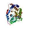
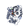
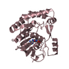
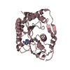
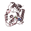
- PDB-3emd: Wesselsbron virus Methyltransferase in complex with Sinefungin an... -
+ Open data

- Basic information
| Entry | Database: PDB / ID: 3emd | ||||||
|---|---|---|---|---|---|---|---|
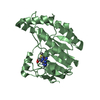
| Title | Wesselsbron virus Methyltransferase in complex with Sinefungin and 7MeGpppA | ||||||
 Components Components | methyltransferase | ||||||
 Keywords Keywords | TRANSFERASE / Flavivirus / RNA capping / Methyltransferase / Guanylyltransferase / viral enzyme structure | ||||||
| Function / homology |  Function and homology information Function and homology informationsymbiont-mediated suppression of host JAK-STAT cascade via inhibition of STAT2 activity / helicase activity / viral capsid / ribonucleoside triphosphate phosphatase activity / double-stranded RNA binding / methyltransferase cap1 / methyltransferase cap1 activity / mRNA 5'-cap (guanine-N7-)-methyltransferase activity / RNA helicase activity / protein dimerization activity ...symbiont-mediated suppression of host JAK-STAT cascade via inhibition of STAT2 activity / helicase activity / viral capsid / ribonucleoside triphosphate phosphatase activity / double-stranded RNA binding / methyltransferase cap1 / methyltransferase cap1 activity / mRNA 5'-cap (guanine-N7-)-methyltransferase activity / RNA helicase activity / protein dimerization activity / symbiont-mediated suppression of host innate immune response / host cell perinuclear region of cytoplasm / host cell endoplasmic reticulum membrane / symbiont-mediated suppression of host type I interferon-mediated signaling pathway / serine-type endopeptidase activity / symbiont-mediated activation of host autophagy / viral RNA genome replication / RNA-directed RNA polymerase activity / fusion of virus membrane with host endosome membrane / symbiont entry into host cell / virion attachment to host cell / host cell nucleus / virion membrane / structural molecule activity / proteolysis / extracellular region / ATP binding / metal ion binding Similarity search - Function | ||||||
| Biological species |  Wesselsbron virus Wesselsbron virus | ||||||
| Method |  X-RAY DIFFRACTION / SYNCHROTRON / MOLECULAR REPLACEMENT / Resolution: 2 Å X-RAY DIFFRACTION / SYNCHROTRON / MOLECULAR REPLACEMENT / Resolution: 2 Å | ||||||
 Authors Authors | Bollati, M. / Milani, M. / Ricagno, S. / Mastrangelo, E. / Bolognesi, M. | ||||||
 Citation Citation | Journal: J.Mol.Biol. / Year: 2009 Title: Recognition of RNA Cap in the Wesselsbron Virus NS5 Methyltransferase Domain: Implications for RNA-Capping Mechanisms in Flavivirus Authors: Bollati, M. / Milani, M. / Mastrangelo, E. / Ricagno, S. / Tedeschi, G. / Nonnis, S. / Decroly, E. / Selisko, B. / de Lamballerie, X. / Coutard, B. / Canard, B. / Bolognesi, M. | ||||||
| History |
|
- Structure visualization
| Structure viewer | Molecule: MolmilJmol/JSmol |
|---|
- Downloads & links
-Download
| PDBx/mmCIF format | 3emd.cif.gz | 78.9 KB | Display | PDBx/mmCIF format |
|---|---|---|---|---|
| PDB format | pdb3emd.ent.gz | 55.3 KB | Display | PDB format |
| PDBx/mmJSON format | 3emd.json.gz | Tree view | PDBx/mmJSON format | |
| Others |  Other downloads Other downloads |
-Validation report
| Arichive directory | https://data.pdbj.org/pub/pdb/validation_reports/em/3emdftp://data.pdbj.org/pub/pdb/validation_reports/em/3emd | HTTPS FTP |
|---|
-Related structure data
| Related structure data |  3eldC  3eluSC 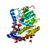 3elwC  3elyC 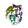 3embC C: citing same article ( S: Starting model for refinement |
|---|---|
| Similar structure data |


-Links
 PDBj
PDBj


- Assembly
| Deposited unit | 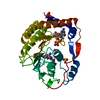
| ||||||||
|---|---|---|---|---|---|---|---|---|---|
| 1 |
| ||||||||
| Unit cell |
|
-Components
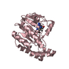
| #1: Protein | Mass: 34193.172 Da / Num. of mol.: 1 Fragment: NS5 N-terminal methyltransferase domain, UNP residues 2500-2791 Source method: isolated from a genetically manipulated source Source: (gene. exp.) Wesselsbron virus / Strain: SAH-177 99871-2 / Plasmid: pDEST14 / Production host:  References: UniProt: C8XPB0, UniProt: D0VX01*PLUS, methyltransferase cap1 | ||||
|---|---|---|---|---|---|
| #2: Chemical | ChemComp-SFG /   Mass: 381.387 Da / Num. of mol.: 1 / Source method: obtained synthetically / Formula: C15H23N7O5 Mass: 381.387 Da / Num. of mol.: 1 / Source method: obtained synthetically / Formula: C15H23N7O5 | ||||
| #3: Chemical | ChemComp-GTA / 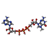  Mass: 787.441 Da / Num. of mol.: 1 / Source method: obtained synthetically / Formula: C21H30N10O17P3 Mass: 787.441 Da / Num. of mol.: 1 / Source method: obtained synthetically / Formula: C21H30N10O17P3 | ||||
| #4: Chemical |   Mass: 96.063 Da / Num. of mol.: 3 / Source method: obtained synthetically / Formula: SO4 Mass: 96.063 Da / Num. of mol.: 3 / Source method: obtained synthetically / Formula: SO4#5: Water | ChemComp-HOH / |  Mass: 18.015 Da / Num. of mol.: 317 / Source method: isolated from a natural source / Formula: H2O Mass: 18.015 Da / Num. of mol.: 317 / Source method: isolated from a natural source / Formula: H2ONonpolymer details | THE THIRD PHOSPHATE GROUP IS NOT VISIBLE IN THE STRUCTURE AS WELL AS THE ADENOSINE. | |
-Experimental details
-Experiment
| Experiment | Method: X-RAY DIFFRACTION / Number of used crystals: 1 |
|---|
- Sample preparation
| Crystal | Density Matthews: 2.13 Å3/Da / Density % sol: 42.38 % |
|---|---|
| Crystal grow | Temperature: 293 K / Method: vapor diffusion, sitting drop / pH: 6.2 Details: 1mM Sinefungin, 1mM 7MeGpppA, 10% PEG 4000, pH 6.2, VAPOR DIFFUSION, SITTING DROP, temperature 293K |
-Data collection
| Diffraction | Mean temperature: 100 K |
|---|---|
| Diffraction source | Source: SYNCHROTRON / Site: ESRF  / Beamline: ID14-2 / Wavelength: 0.933 Å / Beamline: ID14-2 / Wavelength: 0.933 Å |
| Detector | Type: ADSC QUANTUM 4 / Detector: CCD / Date: Nov 10, 2007 / Details: mirrors |
| Radiation | Monochromator: Diamond (111) Ge (220) / Protocol: SINGLE WAVELENGTH / Monochromatic (M) / Laue (L): M / Scattering type: x-ray |
| Radiation wavelength | Wavelength: 0.933 Å / Relative weight: 1 |
| Reflection | Resolution: 2→44.37 Å / Num. all: 21485 / Num. obs: 20462 / % possible obs: 99.6 % / Observed criterion σ(F): 0 / Observed criterion σ(I): 0 / Redundancy: 3.5 % / Rmerge(I) obs: 0.094 / Net I/σ(I): 11.5 |
| Reflection shell | Resolution: 2→2.11 Å / Redundancy: 3.5 % / Rmerge(I) obs: 0.293 / Mean I/σ(I) obs: 4.6 / Num. unique all: 3093 / % possible all: 99.9 |
- Processing
| Software |
| ||||||||||||||||||||||||||||||||||||||||||||||||||||||||||||||||||||||||||||||||||||||||||
|---|---|---|---|---|---|---|---|---|---|---|---|---|---|---|---|---|---|---|---|---|---|---|---|---|---|---|---|---|---|---|---|---|---|---|---|---|---|---|---|---|---|---|---|---|---|---|---|---|---|---|---|---|---|---|---|---|---|---|---|---|---|---|---|---|---|---|---|---|---|---|---|---|---|---|---|---|---|---|---|---|---|---|---|---|---|---|---|---|---|---|---|
| Refinement | Method to determine structure: MOLECULAR REPLACEMENT Starting model: pdb entry 3ELU Resolution: 2→32.21 Å / Cor.coef. Fo:Fc: 0.944 / Cor.coef. Fo:Fc free: 0.908 / Cross valid method: THROUGHOUT / σ(F): 0 / σ(I): 0 / ESU R: 0.172 / ESU R Free: 0.158 / Stereochemistry target values: MAXIMUM LIKELIHOOD / Details: HYDROGENS HAVE BEEN ADDED IN THE RIDING POSITIONS
| ||||||||||||||||||||||||||||||||||||||||||||||||||||||||||||||||||||||||||||||||||||||||||
| Solvent computation | Ion probe radii: 0.8 Å / Shrinkage radii: 0.8 Å / VDW probe radii: 1.2 Å / Solvent model: MASK | ||||||||||||||||||||||||||||||||||||||||||||||||||||||||||||||||||||||||||||||||||||||||||
| Displacement parameters | Biso mean: 13.475 Å2
| ||||||||||||||||||||||||||||||||||||||||||||||||||||||||||||||||||||||||||||||||||||||||||
| Refinement step | Cycle: LAST / Resolution: 2→32.21 Å
| ||||||||||||||||||||||||||||||||||||||||||||||||||||||||||||||||||||||||||||||||||||||||||
| Refine LS restraints |
| ||||||||||||||||||||||||||||||||||||||||||||||||||||||||||||||||||||||||||||||||||||||||||
| LS refinement shell | Resolution: 2→2.052 Å / Total num. of bins used: 20
|
