 Movie
Movie Controller

[English] 日本語
 Yorodumi
- PDB-3ek1: Crystal structure of aldehyde dehydrogenase from brucella meliten... -
+ Open data

- Basic information
| Entry | Database: PDB / ID: 3ek1 | ||||||
|---|---|---|---|---|---|---|---|
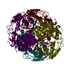
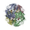
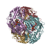
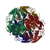
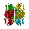
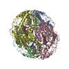
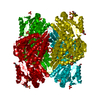
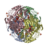
| Title | Crystal structure of aldehyde dehydrogenase from brucella melitensis biovar abortus 2308 | ||||||
 Components Components | Aldehyde dehydrogenase | ||||||
 Keywords Keywords | OXIDOREDUCTASE / SSGCID / ALDEHYDE DEHYDROGENASE / Structural Genomics / Seattle Structural Genomics Center for Infectious Disease | ||||||
| Function / homology |  Function and homology information Function and homology informationsuccinate-semialdehyde dehydrogenase [NAD(P)+] / succinate-semialdehyde dehydrogenase (NAD+) activity / GABA catabolic process / cytosol Similarity search - Function | ||||||
| Biological species |  Brucella melitensis biovar Abortus 2308 (bacteria) Brucella melitensis biovar Abortus 2308 (bacteria) | ||||||
| Method |  X-RAY DIFFRACTION / SYNCHROTRON / molecular replacement, molecular replacement / Resolution: 2.1 Å X-RAY DIFFRACTION / SYNCHROTRON / molecular replacement, molecular replacement / Resolution: 2.1 Å | ||||||
 Authors Authors | Seattle Structural Genomics Center for Infectious Disease (SSGCID) | ||||||
 Citation Citation | Journal: To be Published Title: Crystal structure of aldehyde dehydrogenase from brucella melitensis biovar abortus 2308 Authors: Seattle Structural Genomics Center for Infectious Disease (SSGCID) | ||||||
| History |
|
- Structure visualization
| Structure viewer | Molecule: MolmilJmol/JSmol |
|---|
- Downloads & links
-Download
| PDBx/mmCIF format | 3ek1.cif.gz | 776.9 KB | Display | PDBx/mmCIF format |
|---|---|---|---|---|
| PDB format | pdb3ek1.ent.gz | 634.9 KB | Display | PDB format |
| PDBx/mmJSON format | 3ek1.json.gz | Tree view | PDBx/mmJSON format | |
| Others |  Other downloads Other downloads |
-Validation report
| Arichive directory | https://data.pdbj.org/pub/pdb/validation_reports/ek/3ek1ftp://data.pdbj.org/pub/pdb/validation_reports/ek/3ek1 | HTTPS FTP |
|---|
-Related structure data
| Related structure data | 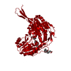 2opxS S: Starting model for refinement |
|---|---|
| Similar structure data | |
| Other databases |


-Links
 PDBj
PDBj

- Assembly
| Deposited unit | 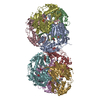
| ||||||||
|---|---|---|---|---|---|---|---|---|---|
| 1 | 
| ||||||||
| 2 | 
| ||||||||
| Unit cell |
|
-Components
| #1: Protein | Mass: 53613.355 Da / Num. of mol.: 8 Source method: isolated from a genetically manipulated source Source: (gene. exp.) Brucella melitensis biovar Abortus 2308 (bacteria)Strain: BIOVAR ABORTUS 2308 / Gene: gabD, BAB1_1655 / Plasmid: PET28-HISSMT / Production host: References: UniProt: Q2YRI3, succinate-semialdehyde dehydrogenase [NAD(P)+] #2: Chemical | ChemComp-SO4 /   Mass: 96.063 Da / Num. of mol.: 8 / Source method: obtained synthetically / Formula: SO4 Mass: 96.063 Da / Num. of mol.: 8 / Source method: obtained synthetically / Formula: SO4#3: Chemical | ChemComp-MES /   Mass: 195.237 Da / Num. of mol.: 8 / Source method: obtained synthetically / Formula: C6H13NO4S / Comment: pH buffer*YM Mass: 195.237 Da / Num. of mol.: 8 / Source method: obtained synthetically / Formula: C6H13NO4S / Comment: pH buffer*YM#4: Water | ChemComp-HOH / |  Mass: 18.015 Da / Num. of mol.: 3142 / Source method: isolated from a natural source / Formula: H2O Mass: 18.015 Da / Num. of mol.: 3142 / Source method: isolated from a natural source / Formula: H2O |
|---|
-Experimental details
-Experiment
| Experiment | Method: X-RAY DIFFRACTION / Number of used crystals: 1 |
|---|
- Sample preparation
| Crystal | Density Matthews: 2.54 Å3/Da / Density % sol: 51.58 % |
|---|---|
| Crystal grow | Temperature: 298 K / Method: vapor diffusion, sitting drop / pH: 6.5 Details: PROPLEX-96 F9: 1.0M AMMONIUM SULPHATE, 100MM MES PH 6.5, VAPOR DIFFUSION, TEMPERATURE 298K, pH 6.50, VAPOR DIFFUSION, SITTING DROP |
-Data collection
| Diffraction | Mean temperature: 100 K |
|---|---|
| Diffraction source | Source: SYNCHROTRON / Site: ALS  / Beamline: 5.0.2 / Wavelength: 0.9999 / Wavelength: 0.9999 Å / Beamline: 5.0.2 / Wavelength: 0.9999 / Wavelength: 0.9999 Å |
| Detector | Type: ADSC QUANTUM 315 / Detector: CCD / Date: Aug 20, 2008 |
| Radiation | Protocol: SINGLE WAVELENGTH / Monochromatic (M) / Laue (L): M / Scattering type: x-ray |
| Radiation wavelength | Wavelength: 0.9999 Å / Relative weight: 1 |
| Reflection | Resolution: 2.1→50 Å / Num. all: 246609 / Num. obs: 231828 / % possible obs: 94 % / Observed criterion σ(F): 0 / Observed criterion σ(I): -3 / Redundancy: 1.81 % / Biso Wilson estimate: 30.6 Å2 / Rmerge(I) obs: 0.061 / Rsym value: 0.061 / Net I/σ(I): 11.02 |
| Reflection shell | Resolution: 2.1→2.15 Å / Redundancy: 1.71 % / Rmerge(I) obs: 0.354 / Mean I/σ(I) obs: 2.3 / Num. unique all: 16866 / Rsym value: 0.354 / % possible all: 92.5 |
- Processing
| Software |
| |||||||||||||||||||||||||||||||||||||||||||||||||||||||||||||||||||||||||||||||||||||
|---|---|---|---|---|---|---|---|---|---|---|---|---|---|---|---|---|---|---|---|---|---|---|---|---|---|---|---|---|---|---|---|---|---|---|---|---|---|---|---|---|---|---|---|---|---|---|---|---|---|---|---|---|---|---|---|---|---|---|---|---|---|---|---|---|---|---|---|---|---|---|---|---|---|---|---|---|---|---|---|---|---|---|---|---|---|---|
| Refinement | Method to determine structure: molecular replacement, molecular replacement Starting model: pdb entry 2opx, modified Resolution: 2.1→19.78 Å / Cor.coef. Fo:Fc: 0.961 / Cor.coef. Fo:Fc free: 0.925 / SU B: 4.67 / SU ML: 0.123 / Isotropic thermal model: isotropic / Cross valid method: THROUGHOUT / σ(F): 0 / σ(I): 0 / ESU R: 0.208 / ESU R Free: 0.188 / Stereochemistry target values: MAXIMUM LIKELIHOOD / Details: HYDROGENS HAVE BEEN ADDED IN THE
| |||||||||||||||||||||||||||||||||||||||||||||||||||||||||||||||||||||||||||||||||||||
| Solvent computation | Ion probe radii: 0.8 Å / Shrinkage radii: 0.8 Å / VDW probe radii: 1.2 Å / Solvent model: MASK | |||||||||||||||||||||||||||||||||||||||||||||||||||||||||||||||||||||||||||||||||||||
| Displacement parameters | Biso mean: 19.83 Å2
| |||||||||||||||||||||||||||||||||||||||||||||||||||||||||||||||||||||||||||||||||||||
| Refinement step | Cycle: LAST / Resolution: 2.1→19.78 Å
| |||||||||||||||||||||||||||||||||||||||||||||||||||||||||||||||||||||||||||||||||||||
| Refine LS restraints |
| |||||||||||||||||||||||||||||||||||||||||||||||||||||||||||||||||||||||||||||||||||||
| LS refinement shell | Resolution: 2.1→2.15 Å / Total num. of bins used: 20
|
