 Movie
Movie Controller

[English] 日本語
 Yorodumi
- PDB-3eiv: Crystal Structure of Single-stranded DNA-binding protein from Str... -
+ Open data

- Basic information
| Entry | Database: PDB / ID: 3eiv | ||||||
|---|---|---|---|---|---|---|---|
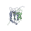
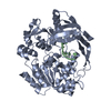
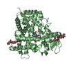
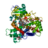
| Title | Crystal Structure of Single-stranded DNA-binding protein from Streptomyces coelicolor | ||||||
 Components Components | Single-stranded DNA-binding protein 2 | ||||||
 Keywords Keywords | DNA BINDING PROTEIN / Single-stranded DNA-binding protein / Streptomyces Coelicolor / DNA damage / DNA repair / DNA replication / DNA-binding / Phosphoprotein | ||||||
| Function / homology |  Function and homology information Function and homology informationnucleoid / enzyme activator activity / single-stranded DNA binding / DNA replication Similarity search - Function | ||||||
| Biological species |  Streptomyces coelicolor (bacteria) Streptomyces coelicolor (bacteria) | ||||||
| Method |  X-RAY DIFFRACTION / SYNCHROTRON / molecular replacement, molecular replacement / molecular replacement / Resolution: 2.141 Å X-RAY DIFFRACTION / SYNCHROTRON / molecular replacement, molecular replacement / molecular replacement / Resolution: 2.141 Å | ||||||
 Authors Authors | Luic, M. / Stefanic, Z. / Vujaklija, D. | ||||||
 Citation Citation | Journal: Acta Crystallogr.,Sect.D / Year: 2009 Title: Structure of the single-stranded DNA-binding protein from Streptomyces coelicolor. Authors: Stefanic, Z. / Vujaklija, D. / Luic, M. | ||||||
| History |
|
- Structure visualization
| Structure viewer | Molecule: MolmilJmol/JSmol |
|---|
- Downloads & links
-Download
| PDBx/mmCIF format | 3eiv.cif.gz | 97.8 KB | Display | PDBx/mmCIF format |
|---|---|---|---|---|
| PDB format | pdb3eiv.ent.gz | 74 KB | Display | PDB format |
| PDBx/mmJSON format | 3eiv.json.gz | Tree view | PDBx/mmJSON format | |
| Others |  Other downloads Other downloads |
-Validation report
| Arichive directory | https://data.pdbj.org/pub/pdb/validation_reports/ei/3eivftp://data.pdbj.org/pub/pdb/validation_reports/ei/3eiv | HTTPS FTP |
|---|
-Related structure data
| Related structure data |  1x3eS S: Starting model for refinement |
|---|---|
| Similar structure data |


-Links
 PDBj
PDBj- Assembly
| Deposited unit | 
| ||||||||||||||||||
|---|---|---|---|---|---|---|---|---|---|---|---|---|---|---|---|---|---|---|---|
| 1 |
| ||||||||||||||||||
| 2 | 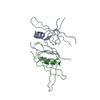
| ||||||||||||||||||
| 3 | 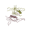
| ||||||||||||||||||
| Unit cell |
| ||||||||||||||||||
| Components on special symmetry positions |
| ||||||||||||||||||
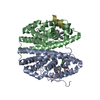
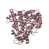
| Details | Biological unit is a tetramer. Biological unit is the same as asym. unit |
-Components
| #1: Protein | Mass: 19925.531 Da / Num. of mol.: 4 / Source method: isolated from a natural source / Source: (natural) Streptomyces coelicolor (bacteria) / References: UniProt: Q9X8U3#2: Water | ChemComp-HOH / |  Mass: 18.015 Da / Num. of mol.: 288 / Source method: isolated from a natural source / Formula: H2O Mass: 18.015 Da / Num. of mol.: 288 / Source method: isolated from a natural source / Formula: H2O |
|---|
-Experimental details
-Experiment
| Experiment | Method: X-RAY DIFFRACTION / Number of used crystals: 1 |
|---|
- Sample preparation
| Crystal | Density Matthews: 2.72 Å3/Da / Density % sol: 54.83 % |
|---|---|
| Crystal grow | Temperature: 291 K / Method: vapor diffusion, hanging drop / pH: 7 Details: 50 mM TRIS-HCl, pH 7.0, VAPOR DIFFUSION, HANGING DROP, temperature 291K |
-Data collection
| Diffraction | Mean temperature: 100 K |
|---|---|
| Diffraction source | Source: SYNCHROTRON / Site: APS  / Beamline: 19-ID / Wavelength: 0.97 Å / Beamline: 19-ID / Wavelength: 0.97 Å |
| Detector | Type: MAR CCD 165 mm / Detector: CCD / Date: Jul 21, 2006 |
| Radiation | Protocol: SINGLE WAVELENGTH / Monochromatic (M) / Laue (L): M / Scattering type: x-ray |
| Radiation wavelength | Wavelength: 0.97 Å / Relative weight: 1 |
| Reflection | Resolution: 2.14→32.2 Å / Num. all: 48150 / Num. obs: 31640 / % possible obs: 65.7 % / Observed criterion σ(F): 3 / Observed criterion σ(I): 3 / Redundancy: 2.44 % / Biso Wilson estimate: 48.39 Å2 / Rmerge(I) obs: 0.038 / Rsym value: 0.0739 / Net I/σ(I): 13.373 |
| Reflection shell | Resolution: 2.14→2.22 Å / Redundancy: 2.48 % / Rmerge(I) obs: 0.35 / Mean I/σ(I) obs: 1.5 / Num. measured all: 22946 / Num. unique all: 5720 / Rsym value: 0.41 / % possible all: 100 |
-Phasing
| Phasing | Method: molecular replacement | |||||||||
|---|---|---|---|---|---|---|---|---|---|---|
| Phasing MR | Model details: Phaser MODE: MR_AUTO
|
- Processing
| Software |
| ||||||||||||||||||||||||||||
|---|---|---|---|---|---|---|---|---|---|---|---|---|---|---|---|---|---|---|---|---|---|---|---|---|---|---|---|---|---|
| Refinement | Method to determine structure: molecular replacement, molecular replacement Starting model: PDB ENTRY 1X3E Resolution: 2.141→31.182 Å / FOM work R set: 0.772 / Cross valid method: THROUGHOUT / σ(F): 2 / σ(I): 2 / Stereochemistry target values: ML
| ||||||||||||||||||||||||||||
| Solvent computation | Shrinkage radii: 0.9 Å / Solvent model: FLAT BULK SOLVENT MODEL / Bsol: 68.116 Å2 / ksol: 0.398 e/Å3 | ||||||||||||||||||||||||||||
| Displacement parameters | Biso max: 90.21 Å2 / Biso mean: 46.76 Å2 / Biso min: 24.89 Å2
| ||||||||||||||||||||||||||||
| Refinement step | Cycle: LAST / Resolution: 2.141→31.182 Å
| ||||||||||||||||||||||||||||
| Refine LS restraints |
|
