 Movie
Movie Controller

[English] 日本語
 Yorodumi
- PDB-3efe: The crystal structure of the thiJ/pfpI family protein from Bacill... -
+ Open data

- Basic information
| Entry | Database: PDB / ID: 3efe | ||||||
|---|---|---|---|---|---|---|---|
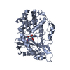
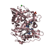
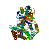
| Title | The crystal structure of the thiJ/pfpI family protein from Bacillus anthracis | ||||||
 Components Components | ThiJ/pfpI family protein | ||||||
 Keywords Keywords | CHAPERONE / thiJ/pfpI family protein / Bacillus anthracis / structural genomics / CSGID / Center for Structural Genomics of Infectious Diseases | ||||||
| Function / homology |  Function and homology information Function and homology information | ||||||
| Biological species |  | ||||||
| Method |  X-RAY DIFFRACTION / SYNCHROTRON / SAD / Resolution: 2.3 Å X-RAY DIFFRACTION / SYNCHROTRON / SAD / Resolution: 2.3 Å | ||||||
 Authors Authors | Zhang, R. / Xu, X. / Cui, H. / Savchenko, A. / Edwards, A. / Anderson, W. / Joachimiak, A. / Center for Structural Genomics of Infectious Diseases (CSGID) | ||||||
 Citation Citation | Journal: To be Published Title: The crystal structure of the thiJ/pfpI family protein from Bacillus anthracis Authors: Zhang, R. / Xu, X. / Cui, H. / Savchenko, A. / Edwards, A. / Anderson, W. / Joachimiak, A. | ||||||
| History |
|
- Structure visualization
| Structure viewer | Molecule: MolmilJmol/JSmol |
|---|
- Downloads & links
-Download
| PDBx/mmCIF format | 3efe.cif.gz | 250.1 KB | Display | PDBx/mmCIF format |
|---|---|---|---|---|
| PDB format | pdb3efe.ent.gz | 204.3 KB | Display | PDB format |
| PDBx/mmJSON format | 3efe.json.gz | Tree view | PDBx/mmJSON format | |
| Others |  Other downloads Other downloads |
-Validation report
| Arichive directory | https://data.pdbj.org/pub/pdb/validation_reports/ef/3efeftp://data.pdbj.org/pub/pdb/validation_reports/ef/3efe | HTTPS FTP |
|---|
-Related structure data
| Similar structure data | |
|---|---|
| Other databases |


-Links
 PDBj
PDBj


- Assembly
| Deposited unit | 
| ||||||||
|---|---|---|---|---|---|---|---|---|---|
| 1 | 
| ||||||||
| 2 | 
| ||||||||
| 3 | 
| ||||||||
| Unit cell |
|
-Components
| #1: Protein | Mass: 23783.445 Da / Num. of mol.: 6 Source method: isolated from a genetically manipulated source Source: (gene. exp.) #2: Chemical | ChemComp-SO4 / |   Mass: 96.063 Da / Num. of mol.: 1 / Source method: obtained synthetically / Formula: SO4 Mass: 96.063 Da / Num. of mol.: 1 / Source method: obtained synthetically / Formula: SO4#3: Water | ChemComp-HOH / |  Mass: 18.015 Da / Num. of mol.: 244 / Source method: isolated from a natural source / Formula: H2O Mass: 18.015 Da / Num. of mol.: 244 / Source method: isolated from a natural source / Formula: H2OSequence details | THE TARGET ID IDP01712 DOES NOT EXIST IN TARGETDB AT THE TIME OF PROCESSING | |
|---|
-Experimental details
-Experiment
| Experiment | Method: X-RAY DIFFRACTION / Number of used crystals: 1 |
|---|
- Sample preparation
| Crystal | Density Matthews: 2.28 Å3/Da / Density % sol: 46.11 % |
|---|---|
| Crystal grow | Temperature: 289 K / Method: vapor diffusion, hanging drop / pH: 7.5 Details: 0.1M Heoes, 0.2M NH4(OAC0), 25% PEG3350, pH 7.5, VAPOR DIFFUSION, HANGING DROP, temperature 289K |
-Data collection
| Diffraction | Mean temperature: 100 K |
|---|---|
| Diffraction source | Source: SYNCHROTRON / Site: APS  / Beamline: 19-BM / Wavelength: 0.9794 Å / Beamline: 19-BM / Wavelength: 0.9794 Å |
| Detector | Type: ADSC QUANTUM 315 / Detector: CCD / Date: Jun 25, 2008 / Details: mirrors |
| Radiation | Monochromator: Si 111 channel / Protocol: SINGLE WAVELENGTH / Monochromatic (M) / Laue (L): M / Scattering type: x-ray |
| Radiation wavelength | Wavelength: 0.9794 Å / Relative weight: 1 |
| Reflection | Resolution: 2.3→83.62 Å / Num. all: 55865 / Num. obs: 54826 / % possible obs: 98.14 % / Observed criterion σ(F): 2 / Observed criterion σ(I): 2 / Redundancy: 8.5 % / Biso Wilson estimate: 39 Å2 / Rmerge(I) obs: 0.118 / Net I/σ(I): 17.5 |
| Reflection shell | Resolution: 2.3→2.359 Å / Redundancy: 6.3 % / Rmerge(I) obs: 0.644 / Mean I/σ(I) obs: 1.65 / Num. unique all: 4303 / % possible all: 83.29 |
- Processing
| Software |
| |||||||||||||||||||||||||||||||||||||||||||||||||||||||||||||||||||||||||||||||||||||||||||||||||||||||||||||||||||||||||||||
|---|---|---|---|---|---|---|---|---|---|---|---|---|---|---|---|---|---|---|---|---|---|---|---|---|---|---|---|---|---|---|---|---|---|---|---|---|---|---|---|---|---|---|---|---|---|---|---|---|---|---|---|---|---|---|---|---|---|---|---|---|---|---|---|---|---|---|---|---|---|---|---|---|---|---|---|---|---|---|---|---|---|---|---|---|---|---|---|---|---|---|---|---|---|---|---|---|---|---|---|---|---|---|---|---|---|---|---|---|---|---|---|---|---|---|---|---|---|---|---|---|---|---|---|---|---|---|
| Refinement | Method to determine structure: SAD / Resolution: 2.3→83.62 Å / Cor.coef. Fo:Fc: 0.958 / Cor.coef. Fo:Fc free: 0.934 / SU B: 16.854 / SU ML: 0.198 / TLS residual ADP flag: LIKELY RESIDUAL / Cross valid method: THROUGHOUT / σ(F): 0 / σ(I): 0 / ESU R: 0.374 / ESU R Free: 0.239 / Stereochemistry target values: MAXIMUM LIKELIHOOD / Details: HYDROGENS HAVE BEEN ADDED IN THE RIDING POSITIONS
| |||||||||||||||||||||||||||||||||||||||||||||||||||||||||||||||||||||||||||||||||||||||||||||||||||||||||||||||||||||||||||||
| Solvent computation | Ion probe radii: 0.8 Å / Shrinkage radii: 0.8 Å / VDW probe radii: 1.2 Å / Solvent model: MASK | |||||||||||||||||||||||||||||||||||||||||||||||||||||||||||||||||||||||||||||||||||||||||||||||||||||||||||||||||||||||||||||
| Displacement parameters | Biso mean: 38.249 Å2
| |||||||||||||||||||||||||||||||||||||||||||||||||||||||||||||||||||||||||||||||||||||||||||||||||||||||||||||||||||||||||||||
| Refinement step | Cycle: LAST / Resolution: 2.3→83.62 Å
| |||||||||||||||||||||||||||||||||||||||||||||||||||||||||||||||||||||||||||||||||||||||||||||||||||||||||||||||||||||||||||||
| Refine LS restraints |
| |||||||||||||||||||||||||||||||||||||||||||||||||||||||||||||||||||||||||||||||||||||||||||||||||||||||||||||||||||||||||||||
| LS refinement shell | Resolution: 2.299→2.359 Å / Total num. of bins used: 20
| |||||||||||||||||||||||||||||||||||||||||||||||||||||||||||||||||||||||||||||||||||||||||||||||||||||||||||||||||||||||||||||
| Refinement TLS params. | Method: refined / Origin x: 83.497 Å / Origin y: 3.89 Å / Origin z: 25.68 Å
| |||||||||||||||||||||||||||||||||||||||||||||||||||||||||||||||||||||||||||||||||||||||||||||||||||||||||||||||||||||||||||||
| Refinement TLS group |
|
