 Movie
Movie Controller

[English] 日本語
 Yorodumi
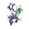
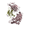
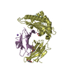
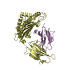
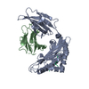
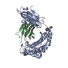
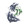
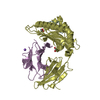
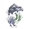
- PDB-3ecb: Crystal structure of mouse H-2Dd in complex with peptide P18-I10 ... -
+ Open data

- Basic information
| Entry | Database: PDB / ID: 3ecb | ||||||
|---|---|---|---|---|---|---|---|
| Title | Crystal structure of mouse H-2Dd in complex with peptide P18-I10 derived from human immunodeficiency virus envelope glycoprotein 120 | ||||||
 Components Components |
| ||||||
 Keywords Keywords | IMMUNE SYSTEM / class I major histompatibility complex / MHC-I / H-2Dd / Glycoprotein / Immune response / Membrane / MHC I / Phosphoprotein / Transmembrane / Immunoglobulin domain / Secreted / AIDS / Apoptosis / Cell membrane / Cleavage on pair of basic residues / Coiled coil / Envelope protein / Fusion protein / Host-virus interaction / Lipoprotein / Palmitate / Viral immunoevasion / Virion | ||||||
| Function / homology |  Function and homology information Function and homology informationEndosomal/Vacuolar pathway / DAP12 interactions / Antigen Presentation: Folding, assembly and peptide loading of class I MHC / ER-Phagosome pathway / DAP12 signaling / Immunoregulatory interactions between a Lymphoid and a non-Lymphoid cell / Dectin-2 family / regulation of membrane depolarization / antigen processing and presentation of peptide antigen via MHC class I / cellular defense response ...Endosomal/Vacuolar pathway / DAP12 interactions / Antigen Presentation: Folding, assembly and peptide loading of class I MHC / ER-Phagosome pathway / DAP12 signaling / Immunoregulatory interactions between a Lymphoid and a non-Lymphoid cell / Dectin-2 family / regulation of membrane depolarization / antigen processing and presentation of peptide antigen via MHC class I / cellular defense response / symbiont-mediated perturbation of host defense response / positive regulation of plasma membrane raft polarization / positive regulation of receptor clustering / Neutrophil degranulation / host cell endosome membrane / lumenal side of endoplasmic reticulum membrane / regulation of iron ion transport / cellular response to iron(III) ion / negative regulation of iron ion transport / negative regulation of forebrain neuron differentiation / antigen processing and presentation of exogenous protein antigen via MHC class Ib, TAP-dependent / iron ion transport / peptide antigen assembly with MHC class I protein complex / regulation of erythrocyte differentiation / response to molecule of bacterial origin / HFE-transferrin receptor complex / MHC class I peptide loading complex / transferrin transport / cellular response to iron ion / negative regulation of receptor-mediated endocytosis / positive regulation of T cell cytokine production / antigen processing and presentation of endogenous peptide antigen via MHC class I / MHC class I protein complex / peptide antigen assembly with MHC class II protein complex / negative regulation of neurogenesis / cellular response to nicotine / MHC class II protein complex / positive regulation of receptor-mediated endocytosis / multicellular organismal-level iron ion homeostasis / positive regulation of T cell mediated cytotoxicity / antigen processing and presentation of exogenous peptide antigen via MHC class II / positive regulation of immune response / peptide antigen binding / phagocytic vesicle membrane / negative regulation of epithelial cell proliferation / positive regulation of T cell activation / sensory perception of smell / positive regulation of cellular senescence / MHC class II protein complex binding / T cell differentiation in thymus / late endosome membrane / antimicrobial humoral immune response mediated by antimicrobial peptide / negative regulation of neuron projection development / antibacterial humoral response / protein refolding / cellular response to lipopolysaccharide / clathrin-dependent endocytosis of virus by host cell / amyloid fibril formation / protein homotetramerization / defense response to Gram-negative bacterium / intracellular iron ion homeostasis / learning or memory / defense response to Gram-positive bacterium / immune response / viral protein processing / fusion of virus membrane with host plasma membrane / external side of plasma membrane / innate immune response / lysosomal membrane / fusion of virus membrane with host endosome membrane / viral envelope / virion attachment to host cell / host cell plasma membrane / virion membrane / structural molecule activity / Golgi apparatus / protein homodimerization activity / : / membrane / identical protein binding / plasma membrane / cytosol Similarity search - Function | ||||||
| Biological species |  | ||||||
| Method |  X-RAY DIFFRACTION / SYNCHROTRON / MOLECULAR REPLACEMENT / Resolution: 1.698 Å X-RAY DIFFRACTION / SYNCHROTRON / MOLECULAR REPLACEMENT / Resolution: 1.698 Å | ||||||
 Authors Authors | Natarajan, K. / Wang, R. / Margulies, D.H. | ||||||
 Citation Citation | Journal: J.Immunol. / Year: 2009 Title: Structural basis of the CD8alphabeta/MHC class i interaction: focused recognition orients CD8beta to a T cell proximal position Authors: Wang, R. / Natarajan, K. / Margulies, D.H. | ||||||
| History |
| ||||||
| Remark 40 | MOLPROBITY STRUCTURE VALIDATION PROGRAMS : MOLPROBITY (KING, REDUCE, AND PROBE) AUTHORS : I.W. ... MOLPROBITY STRUCTURE VALIDATION PROGRAMS : MOLPROBITY (KING, REDUCE, AND PROBE) AUTHORS : I.W.DAVIS,V.B.CHEN, : R.M.IMMORMINO,J.J.HEADD,W.B.ARENDALL,J.M.WORD |
- Structure visualization
| Structure viewer | Molecule: MolmilJmol/JSmol |
|---|
- Downloads & links
-Download
| PDBx/mmCIF format | 3ecb.cif.gz | 177.2 KB | Display | PDBx/mmCIF format |
|---|---|---|---|---|
| PDB format | pdb3ecb.ent.gz | 139.9 KB | Display | PDB format |
| PDBx/mmJSON format | 3ecb.json.gz | Tree view | PDBx/mmJSON format | |
| Others |  Other downloads Other downloads |
-Validation report
| Arichive directory | https://data.pdbj.org/pub/pdb/validation_reports/ec/3ecbftp://data.pdbj.org/pub/pdb/validation_reports/ec/3ecb | HTTPS FTP |
|---|
-Related structure data
| Related structure data |  3dmmC 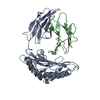 1ddhS C: citing same article ( S: Starting model for refinement |
|---|---|
| Similar structure data |


-Links
 PDBj
PDBj


- Assembly
| Deposited unit | 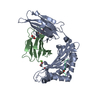
| ||||||||
|---|---|---|---|---|---|---|---|---|---|
| 1 |
| ||||||||
| Unit cell |
|
-Components
-Protein , 2 types, 2 molecules AB
| #1: Protein | Mass: 32265.902 Da / Num. of mol.: 1 / Fragment: UNP residues 26 to 298 Source method: isolated from a genetically manipulated source Source: (gene. exp.)  |
|---|---|
| #2: Protein | Mass: 11791.545 Da / Num. of mol.: 1 / Fragment: UNP residues 21 to 119 Source method: isolated from a genetically manipulated source Source: (gene. exp.) |
-Protein/peptide , 1 types, 1 molecules P
| #3: Protein/peptide | Mass: 1075.265 Da / Num. of mol.: 1 / Fragment: UNP residues 309 to 318 / Source method: obtained synthetically Details: The peptide is chemically synthesized. It is found naturally in Human immunodeficiency virus envelope glycoprotein gp120. References: UniProt: P19551 |
|---|
-Non-polymers , 3 types, 322 molecules 


| #4: Chemical | ChemComp-EDO /  Mass: 62.068 Da / Num. of mol.: 6 / Source method: obtained synthetically / Formula: C2H6O2 Mass: 62.068 Da / Num. of mol.: 6 / Source method: obtained synthetically / Formula: C2H6O2#5: Chemical | ChemComp-MG / |  Mass: 24.305 Da / Num. of mol.: 1 / Source method: obtained synthetically / Formula: Mg Mass: 24.305 Da / Num. of mol.: 1 / Source method: obtained synthetically / Formula: Mg#6: Water | ChemComp-HOH / | Mass: 18.015 Da / Num. of mol.: 315 / Source method: isolated from a natural source / Formula: H2O |
|---|
-Details
| Has protein modification | Y |
|---|
-Experimental details
-Experiment
| Experiment | Method: X-RAY DIFFRACTION / Number of used crystals: 1 |
|---|
- Sample preparation
| Crystal | Density Matthews: 2.58 Å3/Da / Density % sol: 52.29 % |
|---|---|
| Crystal grow | Temperature: 291 K / Method: vapor diffusion, hanging drop / pH: 8 Details: 16% PEG 3350, 0.2M Magnesium formate, , pH 8, VAPOR DIFFUSION, HANGING DROP, temperature 291K |
-Data collection
| Diffraction | Mean temperature: 100 K |
|---|---|
| Diffraction source | Source: SYNCHROTRON / Site: NSLS  / Beamline: X29A / Wavelength: 1.0809 Å / Beamline: X29A / Wavelength: 1.0809 Å |
| Detector | Date: Jul 10, 2008 |
| Radiation | Monochromator: Si 111 / Protocol: SINGLE WAVELENGTH / Monochromatic (M) / Laue (L): M / Scattering type: x-ray |
| Radiation wavelength | Wavelength: 1.0809 Å / Relative weight: 1 |
| Reflection | Resolution: 1.7→30 Å / Num. obs: 51683 / Observed criterion σ(I): 2 / Rmerge(I) obs: 0.06 |
| Reflection shell | Resolution: 1.7→1.76 Å / Redundancy: 10.8 % / Rmerge(I) obs: 0.44 / Mean I/σ(I) obs: 3.7 |
- Processing
| Software |
| |||||||||||||||||||||||||||||||||||||||||||||||||||||||||||||||||||||||||||||||||||||||||||||||||||||||||||||||||||||||||||||||||||||
|---|---|---|---|---|---|---|---|---|---|---|---|---|---|---|---|---|---|---|---|---|---|---|---|---|---|---|---|---|---|---|---|---|---|---|---|---|---|---|---|---|---|---|---|---|---|---|---|---|---|---|---|---|---|---|---|---|---|---|---|---|---|---|---|---|---|---|---|---|---|---|---|---|---|---|---|---|---|---|---|---|---|---|---|---|---|---|---|---|---|---|---|---|---|---|---|---|---|---|---|---|---|---|---|---|---|---|---|---|---|---|---|---|---|---|---|---|---|---|---|---|---|---|---|---|---|---|---|---|---|---|---|---|---|---|
| Refinement | Method to determine structure: MOLECULAR REPLACEMENT Starting model: PDB entry 1DDH Resolution: 1.698→28.798 Å / SU ML: 0.25 / σ(F): 1.34 / Phase error: 23.94 / Stereochemistry target values: ML
| |||||||||||||||||||||||||||||||||||||||||||||||||||||||||||||||||||||||||||||||||||||||||||||||||||||||||||||||||||||||||||||||||||||
| Solvent computation | Shrinkage radii: 0.9 Å / VDW probe radii: 1.11 Å / Solvent model: FLAT BULK SOLVENT MODEL / Bsol: 50.003 Å2 / ksol: 0.351 e/Å3 | |||||||||||||||||||||||||||||||||||||||||||||||||||||||||||||||||||||||||||||||||||||||||||||||||||||||||||||||||||||||||||||||||||||
| Displacement parameters |
| |||||||||||||||||||||||||||||||||||||||||||||||||||||||||||||||||||||||||||||||||||||||||||||||||||||||||||||||||||||||||||||||||||||
| Refinement step | Cycle: LAST / Resolution: 1.698→28.798 Å
| |||||||||||||||||||||||||||||||||||||||||||||||||||||||||||||||||||||||||||||||||||||||||||||||||||||||||||||||||||||||||||||||||||||
| Refine LS restraints |
| |||||||||||||||||||||||||||||||||||||||||||||||||||||||||||||||||||||||||||||||||||||||||||||||||||||||||||||||||||||||||||||||||||||
| LS refinement shell |
|
