 Movie
Movie Controller


+ Open data

- Basic information
| Entry | Database: PDB / ID: 3e7r | ||||||
|---|---|---|---|---|---|---|---|
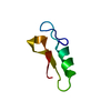
| Title | X-ray Crystal Structure of Racemic Plectasin | ||||||
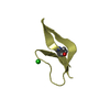
 Components Components | Plectasin | ||||||
 Keywords Keywords | ANTIMICROBIAL PROTEIN / plectasin / racemic protein crystallography / Antibiotic / Antimicrobial / Cleavage on pair of basic residues / Defensin / Secreted | ||||||
| Function / homology |  Function and homology information Function and homology informationpotassium channel regulator activity / toxin activity / defense response to bacterium / host cell plasma membrane / extracellular region Similarity search - Function | ||||||
| Method |  X-RAY DIFFRACTION / SYNCHROTRON / AB INITIO PHASING / Resolution: 1 Å X-RAY DIFFRACTION / SYNCHROTRON / AB INITIO PHASING / Resolution: 1 Å | ||||||
 Authors Authors | Mandal, K. / Pentelute, B.L. / Tereshko, V. / Kossiakoff, A.A. / Kent, S.B.H. | ||||||
 Citation Citation | Journal: Protein Sci. / Year: 2009 Title: Racemic crystallography of synthetic protein enantiomers used to determine the X-ray structure of plectasin by direct methods Authors: Mandal, K. / Pentelute, B.L. / Tereshko, V. / Thammavongsa, V. / Schneewind, O. / Kossiakoff, A.A. / Kent, S.B. | ||||||
| History |
|
- Structure visualization
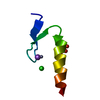
| Structure viewer | Molecule: MolmilJmol/JSmol |
|---|
- Downloads & links
-Download
| PDBx/mmCIF format | 3e7r.cif.gz | 27.3 KB | Display | PDBx/mmCIF format |
|---|---|---|---|---|
| PDB format | pdb3e7r.ent.gz | 18.1 KB | Display | PDB format |
| PDBx/mmJSON format | 3e7r.json.gz | Tree view | PDBx/mmJSON format | |
| Others |  Other downloads Other downloads |
-Validation report
| Arichive directory | https://data.pdbj.org/pub/pdb/validation_reports/e7/3e7rftp://data.pdbj.org/pub/pdb/validation_reports/e7/3e7r | HTTPS FTP |
|---|
-Related structure data


-Links
 PDBj
PDBj

- Assembly
| Deposited unit | 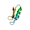
| ||||||||
|---|---|---|---|---|---|---|---|---|---|
| 1 |
| ||||||||
| Unit cell |
|
-Components
| #1: Protein/peptide | Mass: 4415.024 Da / Num. of mol.: 1 / Source method: obtained synthetically Details: The peptide is naturally found in Pseudoplectania nigrella (Ebony cup). References: UniProt: Q53I06 |
|---|---|
| #2: Water | ChemComp-HOH /  Mass: 18.015 Da / Num. of mol.: 24 / Source method: isolated from a natural source / Formula: H2O Mass: 18.015 Da / Num. of mol.: 24 / Source method: isolated from a natural source / Formula: H2O |
| Has protein modification | Y |
-Experimental details
-Experiment
| Experiment | Method: X-RAY DIFFRACTION / Number of used crystals: 1 |
|---|
- Sample preparation
| Crystal | Density Matthews: 1.41 Å3/Da / Density % sol: 14.53 % |
|---|---|
| Crystal grow | Temperature: 292 K / pH: 7 Details: 0.1 M HEPES, 22.5% (w/v) Jeffamine ED-2001, pH 7, VAPOR DIFFUSION, HANGING DROP, temperature 292K |
-Data collection
| Diffraction | Mean temperature: 100 K |
|---|---|
| Diffraction source | Source: SYNCHROTRON / Site: APS  / Beamline: 23-ID-D / Wavelength: 0.97934 / Beamline: 23-ID-D / Wavelength: 0.97934 |
| Detector | Type: MARMOSAIC 300 mm CCD / Detector: CCD / Date: Oct 24, 2007 |
| Radiation | Protocol: SINGLE WAVELENGTH / Monochromatic (M) / Laue (L): M / Scattering type: x-ray |
| Radiation wavelength | Wavelength: 0.97934 Å / Relative weight: 1 |
| Reflection | Resolution: 1→50 Å / Num. obs: 22253 / % possible obs: 83.2 % / Redundancy: 3.5 % / Rmerge(I) obs: 0.068 / Net I/σ(I): 12 |
| Reflection shell | Resolution: 1→1.04 Å / Redundancy: 2.2 % / Rmerge(I) obs: 0.212 / Mean I/σ(I) obs: 4.8 / % possible all: 32 |
- Processing
| Software |
| ||||||||||||||||||||||||||||||||||||||||||||||||||||||||||||||||||||||||||||||||||||||||||||||||||||||||||||||||||||||||||||||||||||||||||||||||||||||||||||||||||||||||||
|---|---|---|---|---|---|---|---|---|---|---|---|---|---|---|---|---|---|---|---|---|---|---|---|---|---|---|---|---|---|---|---|---|---|---|---|---|---|---|---|---|---|---|---|---|---|---|---|---|---|---|---|---|---|---|---|---|---|---|---|---|---|---|---|---|---|---|---|---|---|---|---|---|---|---|---|---|---|---|---|---|---|---|---|---|---|---|---|---|---|---|---|---|---|---|---|---|---|---|---|---|---|---|---|---|---|---|---|---|---|---|---|---|---|---|---|---|---|---|---|---|---|---|---|---|---|---|---|---|---|---|---|---|---|---|---|---|---|---|---|---|---|---|---|---|---|---|---|---|---|---|---|---|---|---|---|---|---|---|---|---|---|---|---|---|---|---|---|---|---|---|---|
| Refinement | Method to determine structure: AB INITIO PHASING / Resolution: 1→22.84 Å / Cor.coef. Fo:Fc: 0.96 / Cor.coef. Fo:Fc free: 0.955 / SU B: 0.522 / SU ML: 0.013 / Cross valid method: THROUGHOUT / ESU R: 0.03 / ESU R Free: 0.03 / Stereochemistry target values: MAXIMUM LIKELIHOOD / Details: HYDROGENS HAVE BEEN ADDED IN THE RIDING POSITIONS
| ||||||||||||||||||||||||||||||||||||||||||||||||||||||||||||||||||||||||||||||||||||||||||||||||||||||||||||||||||||||||||||||||||||||||||||||||||||||||||||||||||||||||||
| Solvent computation | Ion probe radii: 0.8 Å / Shrinkage radii: 0.8 Å / VDW probe radii: 1.2 Å / Solvent model: MASK | ||||||||||||||||||||||||||||||||||||||||||||||||||||||||||||||||||||||||||||||||||||||||||||||||||||||||||||||||||||||||||||||||||||||||||||||||||||||||||||||||||||||||||
| Displacement parameters | Biso mean: 11.78 Å2
| ||||||||||||||||||||||||||||||||||||||||||||||||||||||||||||||||||||||||||||||||||||||||||||||||||||||||||||||||||||||||||||||||||||||||||||||||||||||||||||||||||||||||||
| Refinement step | Cycle: LAST / Resolution: 1→22.84 Å
| ||||||||||||||||||||||||||||||||||||||||||||||||||||||||||||||||||||||||||||||||||||||||||||||||||||||||||||||||||||||||||||||||||||||||||||||||||||||||||||||||||||||||||
| Refine LS restraints |
| ||||||||||||||||||||||||||||||||||||||||||||||||||||||||||||||||||||||||||||||||||||||||||||||||||||||||||||||||||||||||||||||||||||||||||||||||||||||||||||||||||||||||||
| LS refinement shell | Resolution: 1→1.03 Å / Total num. of bins used: 20
|
