 Movie
Movie Controller

[English] 日本語
 Yorodumi
- PDB-3e6h: MHC CLASS I H-2Dd heavy chain complexed with Beta-2 Microglobulin... -
+ Open data

- Basic information
| Entry | Database: PDB / ID: 3e6h | ||||||
|---|---|---|---|---|---|---|---|
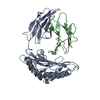
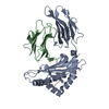
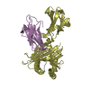
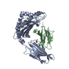
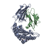
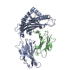
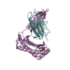
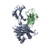
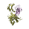
| Title | MHC CLASS I H-2Dd heavy chain complexed with Beta-2 Microglobulin and a variant peptide, PI10, from the human immunodeficiency virus (BaL) envelope glycoprotein 120 | ||||||
 Components Components |
| ||||||
 Keywords Keywords | IMMUNE SYSTEM / COMPLEX (HISTOCOMPATIBILITY-ANTIGEN) / Glycoprotein / Immune response / Membrane / MHC I / Phosphoprotein / Transmembrane / Immunoglobulin domain / Secreted / Envelope protein | ||||||
| Function / homology |  Function and homology information Function and homology informationMHC class Ib protein complex / antigen processing and presentation of exogenous peptide antigen via MHC class Ib / Endosomal/Vacuolar pathway / DAP12 interactions / Antigen Presentation: Folding, assembly and peptide loading of class I MHC / ER-Phagosome pathway / DAP12 signaling / Immunoregulatory interactions between a Lymphoid and a non-Lymphoid cell / Dectin-2 family / regulation of membrane depolarization ...MHC class Ib protein complex / antigen processing and presentation of exogenous peptide antigen via MHC class Ib / Endosomal/Vacuolar pathway / DAP12 interactions / Antigen Presentation: Folding, assembly and peptide loading of class I MHC / ER-Phagosome pathway / DAP12 signaling / Immunoregulatory interactions between a Lymphoid and a non-Lymphoid cell / Dectin-2 family / regulation of membrane depolarization / antigen processing and presentation of peptide antigen via MHC class I / cellular defense response / symbiont-mediated perturbation of host defense response / positive regulation of plasma membrane raft polarization / positive regulation of receptor clustering / Neutrophil degranulation / host cell endosome membrane / lumenal side of endoplasmic reticulum membrane / regulation of iron ion transport / cellular response to iron(III) ion / antigen processing and presentation of exogenous protein antigen via MHC class Ib, TAP-dependent / negative regulation of iron ion transport / negative regulation of forebrain neuron differentiation / regulation of erythrocyte differentiation / iron ion transport / peptide antigen assembly with MHC class I protein complex / response to molecule of bacterial origin / HFE-transferrin receptor complex / MHC class I peptide loading complex / transferrin transport / negative regulation of receptor-mediated endocytosis / cellular response to iron ion / positive regulation of T cell cytokine production / antigen processing and presentation of endogenous peptide antigen via MHC class I / MHC class I protein complex / peptide antigen assembly with MHC class II protein complex / negative regulation of neurogenesis / multicellular organismal-level iron ion homeostasis / cellular response to nicotine / MHC class II protein complex / positive regulation of receptor-mediated endocytosis / positive regulation of T cell mediated cytotoxicity / negative regulation of epithelial cell proliferation / antigen processing and presentation of exogenous peptide antigen via MHC class II / positive regulation of immune response / peptide antigen binding / phagocytic vesicle membrane / positive regulation of T cell activation / sensory perception of smell / positive regulation of cellular senescence / MHC class II protein complex binding / T cell differentiation in thymus / late endosome membrane / negative regulation of neuron projection development / antimicrobial humoral immune response mediated by antimicrobial peptide / antibacterial humoral response / cellular response to lipopolysaccharide / protein refolding / clathrin-dependent endocytosis of virus by host cell / amyloid fibril formation / defense response to Gram-negative bacterium / protein homotetramerization / intracellular iron ion homeostasis / learning or memory / defense response to Gram-positive bacterium / immune response / viral protein processing / fusion of virus membrane with host plasma membrane / external side of plasma membrane / innate immune response / lysosomal membrane / fusion of virus membrane with host endosome membrane / viral envelope / virion attachment to host cell / host cell plasma membrane / virion membrane / structural molecule activity / Golgi apparatus / protein homodimerization activity / : / membrane / identical protein binding / plasma membrane / cytosol Similarity search - Function | ||||||
| Biological species |    Human immunodeficiency virus 1 Human immunodeficiency virus 1 | ||||||
| Method |  X-RAY DIFFRACTION / SYNCHROTRON / MOLECULAR REPLACEMENT / molecular replacement / Resolution: 2.1 Å X-RAY DIFFRACTION / SYNCHROTRON / MOLECULAR REPLACEMENT / molecular replacement / Resolution: 2.1 Å | ||||||
 Authors Authors | Wang, R. / Natarajan, K. / Robinson, H. / Margulies, D.H. | ||||||
 Citation Citation | Journal: J.Immunol. / Year: 2009 Title: Different vaccine vectors delivering the same antigen elicit CD8+ T cell responses with distinct clonotype and epitope specificity Authors: Honda, M. / Wang, R. / Kong, W.P. / Kanekiyo, M. / Akahata, W. / Xu, L. / Matsuo, K. / Natarajan, K. / Robinson, H. / Asher, T.E. / Price, D.A. / Douek, D.C. / Margulies, D.H. / Nabel, G.J. | ||||||
| History |
|
- Structure visualization
| Structure viewer | Molecule: MolmilJmol/JSmol |
|---|
- Downloads & links
-Download
| PDBx/mmCIF format | 3e6h.cif.gz | 94.3 KB | Display | PDBx/mmCIF format |
|---|---|---|---|---|
| PDB format | pdb3e6h.ent.gz | 70.9 KB | Display | PDB format |
| PDBx/mmJSON format | 3e6h.json.gz | Tree view | PDBx/mmJSON format | |
| Others |  Other downloads Other downloads |
-Validation report
| Arichive directory | https://data.pdbj.org/pub/pdb/validation_reports/e6/3e6hftp://data.pdbj.org/pub/pdb/validation_reports/e6/3e6h | HTTPS FTP |
|---|
-Related structure data
| Related structure data | 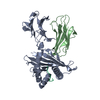 3e6fC  1qo3S S: Starting model for refinement C: citing same article ( |
|---|---|
| Similar structure data |


-Links
 PDBj
PDBj


- Assembly
| Deposited unit | 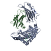
| ||||||||
|---|---|---|---|---|---|---|---|---|---|
| 1 |
| ||||||||
| Unit cell |
|
-Components
| #1: Protein | Mass: 32107.750 Da / Num. of mol.: 1 / Fragment: EXTRACELLULAR DOMAINS Source method: isolated from a genetically manipulated source Source: (gene. exp.)  |
|---|---|
| #2: Protein | Mass: 11791.545 Da / Num. of mol.: 1 / Fragment: EXTRACELLULAR DOMAIN Source method: isolated from a genetically manipulated source Source: (gene. exp.) |
| #3: Protein/peptide | Mass: 1095.272 Da / Num. of mol.: 1 / Source method: obtained synthetically / Details: The peptide was chemically synthesized. / Source: (synth.) Human immunodeficiency virus 1 / References: UniProt: A4L8F7, UniProt: P05877*PLUS |
| #4: Water | ChemComp-HOH /  Mass: 18.015 Da / Num. of mol.: 151 / Source method: isolated from a natural source / Formula: H2O Mass: 18.015 Da / Num. of mol.: 151 / Source method: isolated from a natural source / Formula: H2O |
| Has protein modification | Y |
-Experimental details
-Experiment
| Experiment | Method: X-RAY DIFFRACTION / Number of used crystals: 1 |
|---|
- Sample preparation
| Crystal | Density Matthews: 2.79 Å3/Da / Density % sol: 55.89 % |
|---|---|
| Crystal grow | Temperature: 291 K / Method: vapor diffusion, hanging drop / pH: 6.4 Details: 11% PEG20000,0.1M MES, pH 6.4, VAPOR DIFFUSION, HANGING DROP, temperature 291K |
-Data collection
| Diffraction | Mean temperature: 100 K |
|---|---|
| Diffraction source | Source: SYNCHROTRON / Site: NSLS  / Beamline: X29A / Wavelength: 1 Å / Beamline: X29A / Wavelength: 1 Å |
| Detector | Type: ADSC QUANTUM 4 / Detector: CCD / Date: Jan 1, 2007 |
| Radiation | Protocol: SINGLE WAVELENGTH / Monochromatic (M) / Laue (L): M / Scattering type: x-ray |
| Radiation wavelength | Wavelength: 1 Å / Relative weight: 1 |
| Reflection | Resolution: 2.13→30 Å / Num. all: 30224 / Num. obs: 27990 / % possible obs: 96.8 % / Observed criterion σ(F): 0 / Observed criterion σ(I): -3 / Redundancy: 11.2 % / Rmerge(I) obs: 0.101 / Χ2: 0.9 / Net I/σ(I): 19.3 |
| Reflection shell | Resolution: 2.13→2.21 Å / Redundancy: 3.2 % / Rmerge(I) obs: 0.354 / Mean I/σ(I) obs: 11.9 / Num. unique all: 2087 / Χ2: 0.479 / % possible all: 74.1 |
-Phasing
| Phasing | Method: molecular replacement |
|---|
- Processing
| Software |
| ||||||||||||||||||||||||||||
|---|---|---|---|---|---|---|---|---|---|---|---|---|---|---|---|---|---|---|---|---|---|---|---|---|---|---|---|---|---|
| Refinement | Method to determine structure: MOLECULAR REPLACEMENT Starting model: PDB ENTRY 1QO3 Resolution: 2.1→29.52 Å / Occupancy max: 1 / Occupancy min: 1 / Isotropic thermal model: Isotropic / Cross valid method: THROUGHOUT / σ(F): 0 / Stereochemistry target values: Engh & Huber
| ||||||||||||||||||||||||||||
| Solvent computation | Bsol: 39.346 Å2 | ||||||||||||||||||||||||||||
| Displacement parameters | Biso max: 101.26 Å2 / Biso mean: 27.378 Å2 / Biso min: 7.39 Å2
| ||||||||||||||||||||||||||||
| Refine analyze |
| ||||||||||||||||||||||||||||
| Refinement step | Cycle: LAST / Resolution: 2.1→29.52 Å
| ||||||||||||||||||||||||||||
| Refine LS restraints |
| ||||||||||||||||||||||||||||
| Xplor file |
|
