 Movie
Movie Controller

[English] 日本語
 Yorodumi
- PDB-3e56: The 2.0 Angstrom Resolution Crystal Structure of NpR1517, a Putat... -
+ Open data

- Basic information
| Entry | Database: PDB / ID: 3.0E+56 | ||||||
|---|---|---|---|---|---|---|---|
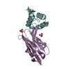
| Title | The 2.0 Angstrom Resolution Crystal Structure of NpR1517, a Putative Heterocyst Differentiation Inhibitor from Nostoc punctiforme | ||||||
 Components Components | Putative uncharacterized protein | ||||||
 Keywords Keywords | UNKNOWN FUNCTION / alpha-beta sandwich / interlocked homodimer / ASR1734 | ||||||
| Function / homology | Ubiquitin-like (UB roll) / Ubiquitin-like (UB roll) - #10 / Npun R1517 / Npun R1517 / Other non-globular / Special / Npun R1517 domain-containing protein Function and homology information Function and homology information | ||||||
| Biological species |  Nostoc punctiforme (bacteria) Nostoc punctiforme (bacteria) | ||||||
| Method |  X-RAY DIFFRACTION / sulfur anomalous scattering / Resolution: 2.01 Å X-RAY DIFFRACTION / sulfur anomalous scattering / Resolution: 2.01 Å | ||||||
 Authors Authors | Kennedy, M.A. / Ni, S. / Smola, M.J. | ||||||
 Citation Citation | Journal: Proteins / Year: 2008 Title: Crystal structure of Npun_R1517, a putative negative regulator of heterocyst differentiation from Nostoc punctiforme PCC 73102. Authors: Ni, S. / Benning, M.M. / Smola, M.J. / Feldmann, E.A. / Kennedy, M.A. | ||||||
| History |
|
- Structure visualization
| Structure viewer | Molecule: MolmilJmol/JSmol |
|---|
- Downloads & links
-Download
| PDBx/mmCIF format | 3e56.cif.gz | 45.2 KB | Display | PDBx/mmCIF format |
|---|---|---|---|---|
| PDB format | pdb3e56.ent.gz | 31.9 KB | Display | PDB format |
| PDBx/mmJSON format | 3e56.json.gz | Tree view | PDBx/mmJSON format | |
| Others |  Other downloads Other downloads |
-Validation report
| Arichive directory | https://data.pdbj.org/pub/pdb/validation_reports/e5/3e56ftp://data.pdbj.org/pub/pdb/validation_reports/e5/3e56 | HTTPS FTP |
|---|
-Related structure data
| Similar structure data |
|---|


-Links
 PDBj
PDBj- Assembly
| Deposited unit | 
| |||||||||
|---|---|---|---|---|---|---|---|---|---|---|
| 1 | 
| |||||||||
| Unit cell |
| |||||||||
| Components on special symmetry positions |
|
-Components
| #1: Protein | Mass: 12916.604 Da / Num. of mol.: 1 / Fragment: UNP residues 1-88 Source method: isolated from a genetically manipulated source Details: cytosolic / Source: (gene. exp.) Nostoc punctiforme (bacteria) / Strain: PCC 73102 / Gene: Npun_R1517 / Plasmid: pET28b / Production host: |
|---|---|
| #2: Water | ChemComp-HOH /  Mass: 18.015 Da / Num. of mol.: 59 / Source method: isolated from a natural source / Formula: H2O Mass: 18.015 Da / Num. of mol.: 59 / Source method: isolated from a natural source / Formula: H2O |
-Experimental details
-Experiment
| Experiment | Method: X-RAY DIFFRACTION / Number of used crystals: 1 |
|---|
- Sample preparation
| Crystal | Density Matthews: 1.74 Å3/Da / Density % sol: 29.11 % |
|---|---|
| Crystal grow | Temperature: 295 K / Method: vapor diffusion, hanging drop / pH: 7.5 Details: .02M HEPES, 20% glycerol, pH 7.5, VAPOR DIFFUSION, HANGING DROP, temperature 295K |
-Data collection
| Diffraction | Mean temperature: 100 K |
|---|---|
| Diffraction source | Source: ROTATING ANODE / Type: BRUKER AXS MICROSTAR / Wavelength: 1.5418 Å |
| Detector | Type: BRUKER SMART 6000 / Detector: CCD / Date: May 15, 2008 / Details: mirrors |
| Radiation | Monochromator: Layered Montel mirrors / Protocol: SINGLE WAVELENGTH / Monochromatic (M) / Laue (L): M / Scattering type: x-ray |
| Radiation wavelength | Wavelength: 1.5418 Å / Relative weight: 1 |
| Reflection | Resolution: 2.01→39.9 Å / Num. all: 6345 / Num. obs: 6345 / % possible obs: 95.9 % / Observed criterion σ(I): 3 / Redundancy: 105.86 % / Biso Wilson estimate: 24.37 Å2 / Rsym value: 0.06 / Net I/σ(I): 76.22 |
| Reflection shell | Resolution: 2.01→2.05 Å / Redundancy: 19.53 % / Mean I/σ(I) obs: 9.15 / Num. unique all: 1604 / Rsym value: 0.28 / % possible all: 95.9 |
- Processing
| Software |
| |||||||||||||||||||||||||||||||||||||||||||||||||||||||||||||||||||||||||||||||||||||||||||||||||||||||||
|---|---|---|---|---|---|---|---|---|---|---|---|---|---|---|---|---|---|---|---|---|---|---|---|---|---|---|---|---|---|---|---|---|---|---|---|---|---|---|---|---|---|---|---|---|---|---|---|---|---|---|---|---|---|---|---|---|---|---|---|---|---|---|---|---|---|---|---|---|---|---|---|---|---|---|---|---|---|---|---|---|---|---|---|---|---|---|---|---|---|---|---|---|---|---|---|---|---|---|---|---|---|---|---|---|---|---|
| Refinement | Method to determine structure: sulfur anomalous scattering / Resolution: 2.01→39.9 Å / Cor.coef. Fo:Fc: 0.953 / Cor.coef. Fo:Fc free: 0.934 / SU B: 8.475 / SU ML: 0.104 / Cross valid method: THROUGHOUT / σ(I): 3 / ESU R: 2.082 / ESU R Free: 0.162 / Stereochemistry target values: MAXIMUM LIKELIHOOD
| |||||||||||||||||||||||||||||||||||||||||||||||||||||||||||||||||||||||||||||||||||||||||||||||||||||||||
| Solvent computation | Ion probe radii: 0.8 Å / Shrinkage radii: 0.8 Å / VDW probe radii: 1.4 Å / Solvent model: MASK | |||||||||||||||||||||||||||||||||||||||||||||||||||||||||||||||||||||||||||||||||||||||||||||||||||||||||
| Displacement parameters | Biso mean: 24.854 Å2
| |||||||||||||||||||||||||||||||||||||||||||||||||||||||||||||||||||||||||||||||||||||||||||||||||||||||||
| Refinement step | Cycle: LAST / Resolution: 2.01→39.9 Å
| |||||||||||||||||||||||||||||||||||||||||||||||||||||||||||||||||||||||||||||||||||||||||||||||||||||||||
| Refine LS restraints |
| |||||||||||||||||||||||||||||||||||||||||||||||||||||||||||||||||||||||||||||||||||||||||||||||||||||||||
| LS refinement shell | Resolution: 2.01→2.063 Å / Total num. of bins used: 20
|
