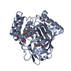
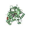
hydrogen peroxide metabolic process / catalase activity / regulation of reactive oxygen species metabolic process / Oxidoreductases; Acting on a peroxide as acceptor; Peroxidases / hydrogen peroxide catabolic process / peroxidase activity / response to hydrogen peroxide / iron ion binding / heme binding / cytoplasm Similarity search - Function
Type: MAR CCD 165 mm / Detector: CCD / Date: Aug 31, 2006 / Details: mirrors
Radiation
Monochromator: Si 111 CHANNEL / Protocol: SINGLE WAVELENGTH / Monochromatic (M) / Laue (L): M / Scattering type: x-ray
Radiation wavelength
Wavelength: 1.38079 Å / Relative weight: 1
Reflection
Resolution: 1.8→36.5 Å / Num. all: 75154 / Num. obs: 75154 / % possible obs: 87.6 % / Observed criterion σ(I): -3 / Redundancy: 2.2 % / Biso Wilson estimate: 23.5 Å2 / Rsym value: 0.057 / Net I/σ(I): 19.5
Reflection shell
Resolution: 1.8→1.86 Å / Redundancy: 2.1 % / Mean I/σ(I) obs: 2 / Num. unique all: 7350 / Rsym value: 0.48 / % possible all: 86.5
-
Processing
Software
Name
Classification
MAR345dtb
datacollection
CNS
refinement
HKL-2000
datareduction
HKL-2000
datascaling
CNS
phasing
Refinement
Method to determine structure: MOLECULAR REPLACEMENT Starting model: Model of lower resolution structure based on Fe MAD data Resolution: 1.8→36.5 Å / Isotropic thermal model: restrained / Cross valid method: THROUGHOUT / Stereochemistry target values: Engh & Huber
Rfactor
Num. reflection
% reflection
Selection details
Rfree
0.21
2090
-
RANDOM
Rwork
0.197
-
-
-
all
-
71376
-
-
obs
-
71376
83.2 %
-
Displacement parameters
Biso mean: 31 Å2
Baniso -1
Baniso -2
Baniso -3
1-
-3.48 Å2
0 Å2
0 Å2
2-
-
4.02 Å2
0 Å2
3-
-
-
-0.54 Å2
Refine analyze
Free
Obs
Luzzati coordinate error
0.22 Å
0.2 Å
Luzzati d res low
-
5 Å
Luzzati sigma a
0.18 Å
0.14 Å
Refinement step
Cycle: LAST / Resolution: 1.8→36.5 Å
Protein
Nucleic acid
Ligand
Solvent
Total
Num. atoms
4539
0
165
367
5071
Refine LS restraints
Refine-ID
Type
Dev ideal
X-RAY DIFFRACTION
c_bond_d
0.005
X-RAY DIFFRACTION
c_angle_deg
1.2
X-RAY DIFFRACTION
c_dihedral_angle_d
23.4
X-RAY DIFFRACTION
c_improper_angle_d
0.74
+
About Yorodumi
-
News
-
Feb 9, 2022. New format data for meta-information of EMDB entries
New format data for meta-information of EMDB entries
Version 3 of the EMDB header file is now the official format.
The previous official version 1.9 will be removed from the archive.
In the structure databanks used in Yorodumi, some data are registered as the other names, "COVID-19 virus" and "2019-nCoV". Here are the details of the virus and the list of structure data.
Jan 31, 2019. EMDB accession codes are about to change! (news from PDBe EMDB page)
EMDB accession codes are about to change! (news from PDBe EMDB page)
The allocation of 4 digits for EMDB accession codes will soon come to an end. Whilst these codes will remain in use, new EMDB accession codes will include an additional digit and will expand incrementally as the available range of codes is exhausted. The current 4-digit format prefixed with “EMD-” (i.e. EMD-XXXX) will advance to a 5-digit format (i.e. EMD-XXXXX), and so on. It is currently estimated that the 4-digit codes will be depleted around Spring 2019, at which point the 5-digit format will come into force.
The EM Navigator/Yorodumi systems omit the EMD- prefix.
Related info.:Q: What is EMD? / ID/Accession-code notation in Yorodumi/EM Navigator
Yorodumi is a browser for structure data from EMDB, PDB, SASBDB, etc.
This page is also the successor to EM Navigator detail page, and also detail information page/front-end page for Omokage search.
The word "yorodu" (or yorozu) is an old Japanese word meaning "ten thousand". "mi" (miru) is to see.
Related info.:EMDB / PDB / SASBDB / Comparison of 3 databanks / Yorodumi Search / Aug 31, 2016. New EM Navigator & Yorodumi / Yorodumi Papers / Jmol/JSmol / Function and homology information / Changes in new EM Navigator and Yorodumi
 Movie
Movie Controller

 Yorodumi
 Open data
 Basic information
 Components
 Keywords
 Function and homology information
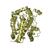
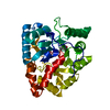
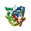
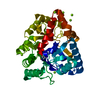
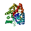
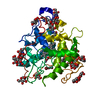
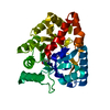
 Mycobacterium avium subsp. paratuberculosis (bacteria)
 X-RAY DIFFRACTION /
  Authors
 Citation
 Structure visualization
 Downloads & links
 Other downloads
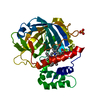


 PDBj
PDBj
 Assembly
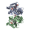


 Mass: 616.487 Da / Num. of mol.: 2 / Source method: obtained synthetically / Formula: C34H32FeN4O4
 Mass: 118.174 Da / Num. of mol.: 6 / Source method: obtained synthetically / Formula: C6H14O2
 Mass: 94.971 Da / Num. of mol.: 5 / Source method: obtained synthetically / Formula: PO4
 Mass: 92.094 Da / Num. of mol.: 1 / Source method: obtained synthetically / Formula: C3H8O3
 Sample preparation
 / Beamline: GCPCC / Wavelength: 1.38079 Å
 Processing