 Movie
Movie Controller


+ Open data

- Basic information
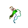
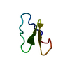
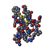
| Entry | Database: PDB / ID: 3e4h | ||||||
|---|---|---|---|---|---|---|---|
| Title | Crystal structure of the cyclotide varv F | ||||||
 Components Components | Varv peptide F,Varv peptide F | ||||||
 Keywords Keywords | PLANT PROTEIN / cyclotide / circular proteins / cystine knot / cyclization / Knottin / Plant defense | ||||||
| Function / homology | Cyclotide, moebius, conserved site / Cyclotides Moebius subfamily signature. / Cyclotides profile. / Cyclotide / Cyclotide superfamily / Cyclotide family / defense response / Varv peptide F Function and homology information Function and homology information | ||||||
| Biological species |  Viola arvensis (European field pansy) Viola arvensis (European field pansy) | ||||||
| Method |  X-RAY DIFFRACTION / SYNCHROTRON / MOLECULAR REPLACEMENT / Resolution: 1.8 Å X-RAY DIFFRACTION / SYNCHROTRON / MOLECULAR REPLACEMENT / Resolution: 1.8 Å | ||||||
 Authors Authors | Hu, S.H. | ||||||
 Citation Citation | Journal: J.Biol.Chem. / Year: 2009 Title: Combined X-ray and NMR Analysis of the Stability of the Cyclotide Cystine Knot Fold That Underpins Its Insecticidal Activity and Potential Use as a Drug Scaffold Authors: Wang, C.K. / Hu, S.H. / Martin, J.L. / Hajdu, J. / Bohlin, L. / Claeson, P. / Rosengren, K.J. / Tang, J. / Tan, N.H. / Craik, D.J. | ||||||
| History |
|
- Structure visualization
| Structure viewer | Molecule: MolmilJmol/JSmol |
|---|
- Downloads & links
-Download
| PDBx/mmCIF format | 3e4h.cif.gz | 16.6 KB | Display | PDBx/mmCIF format |
|---|---|---|---|---|
| PDB format | pdb3e4h.ent.gz | 10.4 KB | Display | PDB format |
| PDBx/mmJSON format | 3e4h.json.gz | Tree view | PDBx/mmJSON format | |
| Others |  Other downloads Other downloads |
-Validation report
| Arichive directory | https://data.pdbj.org/pub/pdb/validation_reports/e4/3e4hftp://data.pdbj.org/pub/pdb/validation_reports/e4/3e4h | HTTPS FTP |
|---|
-Related structure data
| Related structure data |  2k7gC  1nb1S S: Starting model for refinement C: citing same article ( |
|---|---|
| Similar structure data |


-Links
 PDBj
PDBj- Assembly
| Deposited unit | 
| |||||||||
|---|---|---|---|---|---|---|---|---|---|---|
| 1 |
| |||||||||
| 2 | x 6
| |||||||||
| Unit cell |
| |||||||||
| Components on special symmetry positions |
|
-Components
| #1: Protein/peptide | Mass: 2984.432 Da / Num. of mol.: 1 / Source method: isolated from a natural source / Source: (natural) Viola arvensis (European field pansy) / References: UniProt: P58451 |
|---|---|
| #2: Water | ChemComp-HOH /  Mass: 18.015 Da / Num. of mol.: 45 / Source method: isolated from a natural source / Formula: H2O Mass: 18.015 Da / Num. of mol.: 45 / Source method: isolated from a natural source / Formula: H2O |
| Has protein modification | Y |
-Experimental details
-Experiment
| Experiment | Method: X-RAY DIFFRACTION / Number of used crystals: 1 |
|---|
- Sample preparation
| Crystal | Density Matthews: 4.15 Å3/Da / Density % sol: 70.36 % |
|---|---|
| Crystal grow | Temperature: 293 K / Method: vapor diffusion, hanging drop / pH: 9 Details: 2.0M MgCl2, 0.1M bicine, pH9.0, VAPOR DIFFUSION, HANGING DROP, temperature 293K |
-Data collection
| Diffraction | Mean temperature: 100 K |
|---|---|
| Diffraction source | Source: SYNCHROTRON / Site: MAX II  / Beamline: I711 / Wavelength: 1.052 Å / Beamline: I711 / Wavelength: 1.052 Å |
| Detector | Type: MAR CCD 165 mm / Detector: CCD / Date: Jul 6, 2000 / Details: Osmic Max-Flux optics |
| Radiation | Protocol: SINGLE WAVELENGTH / Monochromatic (M) / Laue (L): M / Scattering type: x-ray |
| Radiation wavelength | Wavelength: 1.052 Å / Relative weight: 1 |
| Reflection | Resolution: 1.8→30 Å / Num. obs: 4978 / % possible obs: 99.9 % / Observed criterion σ(I): 0 / Redundancy: 35.8 % / Biso Wilson estimate: 14.2 Å2 / Rmerge(I) obs: 0.061 |
| Reflection shell | Resolution: 1.8→1.86 Å / Redundancy: 35.6 % / Rmerge(I) obs: 0.252 / Mean I/σ(I) obs: 8.9 / Num. unique all: 479 / % possible all: 99.6 |
- Processing
| Software |
| |||||||||||||||||||||||||
|---|---|---|---|---|---|---|---|---|---|---|---|---|---|---|---|---|---|---|---|---|---|---|---|---|---|---|
| Refinement | Method to determine structure: MOLECULAR REPLACEMENT Starting model: PDB ENTRY 1NB1 Resolution: 1.8→30 Å / Isotropic thermal model: Isotropic / Cross valid method: THROUGHOUT / σ(F): 0 / Stereochemistry target values: Engh & Huber
| |||||||||||||||||||||||||
| Displacement parameters | Biso mean: 21 Å2 | |||||||||||||||||||||||||
| Refine analyze |
| |||||||||||||||||||||||||
| Refinement step | Cycle: LAST / Resolution: 1.8→30 Å
| |||||||||||||||||||||||||
| Refine LS restraints |
| |||||||||||||||||||||||||
| LS refinement shell | Resolution: 1.8→1.86 Å / Rfactor Rfree error: 0.015
|
