 Movie
Movie Controller


+ Open data

- Basic information
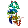
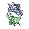
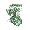
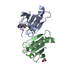
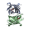
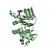
| Entry | Database: PDB / ID: 3e3c | ||||||
|---|---|---|---|---|---|---|---|
| Title | Structure of GrlR-lipid complex | ||||||
 Components Components | L0044 | ||||||
 Keywords Keywords | LIPID BINDING PROTEIN / GrlR / LEE regulator / lipid binding | ||||||
| Function / homology | T3SS negative regulator GrlR / T3SS negative regulator GrlR domain superfamily / Lipocalin / Beta Barrel / Mainly Beta / (2R)-2-HYDROXY-3-(PHOSPHONOOXY)PROPYL HEXANOATE / L0044 / GrlR Function and homology information Function and homology information | ||||||
| Biological species |  | ||||||
| Method |  X-RAY DIFFRACTION / SYNCHROTRON / MOLECULAR REPLACEMENT / Resolution: 2.5 Å X-RAY DIFFRACTION / SYNCHROTRON / MOLECULAR REPLACEMENT / Resolution: 2.5 Å | ||||||
 Authors Authors | Jobichen, C. / Sivaraman, J. | ||||||
 Citation Citation | Journal: Biochem.J. / Year: 2009 Title: Identification and characterization of the lipid binding property of GrlR, a locus of enterocyte effacement regulator. Authors: Jobichen, C. / Fernandis, A.Z. / Velazquez-Campoy, A. / Leung, K.Y. / Mok, Y.K. / Wenk, M.R. / Sivaraman, J. | ||||||
| History |
|
- Structure visualization
| Structure viewer | Molecule: MolmilJmol/JSmol |
|---|
- Downloads & links
-Download
| PDBx/mmCIF format | 3e3c.cif.gz | 64.3 KB | Display | PDBx/mmCIF format |
|---|---|---|---|---|
| PDB format | pdb3e3c.ent.gz | 47.5 KB | Display | PDB format |
| PDBx/mmJSON format | 3e3c.json.gz | Tree view | PDBx/mmJSON format | |
| Others |  Other downloads Other downloads |
-Validation report
| Arichive directory | https://data.pdbj.org/pub/pdb/validation_reports/e3/3e3cftp://data.pdbj.org/pub/pdb/validation_reports/e3/3e3c | HTTPS FTP |
|---|
-Related structure data
| Related structure data |  2ovsS S: Starting model for refinement |
|---|---|
| Similar structure data |


-Links
 PDBj
PDBj- Assembly
| Deposited unit | 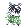
| ||||||||
|---|---|---|---|---|---|---|---|---|---|
| 1 |
| ||||||||
| Unit cell |
|
-Components
| #1: Protein | Mass: 13235.131 Da / Num. of mol.: 2 Source method: isolated from a genetically manipulated source Source: (gene. exp.) #2: Chemical | 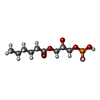  Mass: 270.217 Da / Num. of mol.: 2 / Source method: obtained synthetically / Formula: C9H19O7P Mass: 270.217 Da / Num. of mol.: 2 / Source method: obtained synthetically / Formula: C9H19O7P#3: Water | ChemComp-HOH / |  Mass: 18.015 Da / Num. of mol.: 264 / Source method: isolated from a natural source / Formula: H2O Mass: 18.015 Da / Num. of mol.: 264 / Source method: isolated from a natural source / Formula: H2O |
|---|
-Experimental details
-Experiment
| Experiment | Method: X-RAY DIFFRACTION / Number of used crystals: 1 |
|---|
- Sample preparation
| Crystal | Density Matthews: 2.28 Å3/Da / Density % sol: 45.95 % |
|---|---|
| Crystal grow | Temperature: 298 K / Method: vapor diffusion, hanging drop / pH: 7.5 Details: 25% ethylene Glycol, 4% Tertiary Butanol, 4% triflouroethanol, pH 7.5, VAPOR DIFFUSION, HANGING DROP, temperature 298K |
-Data collection
| Diffraction | Mean temperature: 298 K |
|---|---|
| Diffraction source | Source: SYNCHROTRON / Site: NSLS  / Beamline: X29A / Wavelength: 1.1 Å / Beamline: X29A / Wavelength: 1.1 Å |
| Detector | Type: ADSC QUANTUM 315 / Detector: CCD / Date: Sep 26, 2007 / Details: Mirrors |
| Radiation | Monochromator: SAGITALLY FOCUSED Si(111) / Protocol: SINGLE WAVELENGTH / Monochromatic (M) / Laue (L): M / Scattering type: x-ray |
| Radiation wavelength | Wavelength: 1.1 Å / Relative weight: 1 |
| Reflection | Resolution: 2.5→15 Å / Num. all: 8751 / Num. obs: 8472 / % possible obs: 97 % / Observed criterion σ(F): 2 / Observed criterion σ(I): 2 / Redundancy: 6.7 % / Rmerge(I) obs: 0.103 / Net I/σ(I): 8.2 |
| Reflection shell | Resolution: 2.5→2.59 Å / Redundancy: 4.9 % / Rmerge(I) obs: 0.248 / Num. unique all: 781 / % possible all: 89.9 |
- Processing
| Software |
| |||||||||||||||||||||||||
|---|---|---|---|---|---|---|---|---|---|---|---|---|---|---|---|---|---|---|---|---|---|---|---|---|---|---|
| Refinement | Method to determine structure: MOLECULAR REPLACEMENT Starting model: PDB entry 2OVS Resolution: 2.5→15 Å / Isotropic thermal model: Isotropic / Cross valid method: THROUGHOUT / σ(F): 2 / σ(I): 2 / Stereochemistry target values: Engh & Huber
| |||||||||||||||||||||||||
| Refinement step | Cycle: LAST / Resolution: 2.5→15 Å
| |||||||||||||||||||||||||
| Refine LS restraints |
|
