 Movie
Movie Controller

[English] 日本語
 Yorodumi
- PDB-3e2y: Crystal structure of mouse kynurenine aminotransferase III in com... -
+ Open data

- Basic information
| Entry | Database: PDB / ID: 3e2y | ||||||
|---|---|---|---|---|---|---|---|
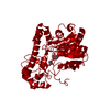
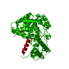
| Title | Crystal structure of mouse kynurenine aminotransferase III in complex with glutamine | ||||||
 Components Components | Kynurenine-oxoglutarate transaminase 3 | ||||||
 Keywords Keywords | TRANSFERASE / LYASE / alpha beta protein / PLP dependent protein / Aminotransferase / Pyridoxal phosphate | ||||||
| Function / homology |  Function and homology information Function and homology information: / kynurenine-glyoxylate transaminase / L-kynurenine:glyoxylate transaminase activity / cysteine-S-conjugate beta-lyase activity / cysteine-S-conjugate beta-lyase / kynurenine-oxoglutarate transaminase / L-kynurenine:2-oxoglutarate transaminase activity / : / 2-oxoglutarate metabolic process / amino acid metabolic process ...: / kynurenine-glyoxylate transaminase / L-kynurenine:glyoxylate transaminase activity / cysteine-S-conjugate beta-lyase activity / cysteine-S-conjugate beta-lyase / kynurenine-oxoglutarate transaminase / L-kynurenine:2-oxoglutarate transaminase activity / : / 2-oxoglutarate metabolic process / amino acid metabolic process / pyridoxal phosphate binding / protein homodimerization activity / mitochondrion / cytoplasm Similarity search - Function | ||||||
| Biological species |  | ||||||
| Method |  X-RAY DIFFRACTION / SYNCHROTRON / MOLECULAR REPLACEMENT / Resolution: 2.26 Å X-RAY DIFFRACTION / SYNCHROTRON / MOLECULAR REPLACEMENT / Resolution: 2.26 Å | ||||||
 Authors Authors | Han, Q. / Robinson, R. / Cai, T. / Tagle, D.A. / Li, J. | ||||||
 Citation Citation | Journal: Mol. Cell. Biol. / Year: 2018 Title: Correction for Han et al., "Biochemical and Structural Properties of Mouse Kynurenine Aminotransferase III". Authors: Han, Q. / Robinson, H. / Cai, T. / Tagle, D.A. / Li, J. #1: Journal: Mol.Cell.Biol. / Year: 2009 Title: Biochemical and structural properties of mouse kynurenine aminotransferase III. Authors: Han, Q. / Robinson, H. / Cai, T. / Tagle, D.A. / Li, J. | ||||||
| History |
|
- Structure visualization
| Structure viewer | Molecule: MolmilJmol/JSmol |
|---|
- Downloads & links
-Download
| PDBx/mmCIF format | 3e2y.cif.gz | 182.1 KB | Display | PDBx/mmCIF format |
|---|---|---|---|---|
| PDB format | pdb3e2y.ent.gz | 143.6 KB | Display | PDB format |
| PDBx/mmJSON format | 3e2y.json.gz | Tree view | PDBx/mmJSON format | |
| Others |  Other downloads Other downloads |
-Validation report
| Arichive directory | https://data.pdbj.org/pub/pdb/validation_reports/e2/3e2yftp://data.pdbj.org/pub/pdb/validation_reports/e2/3e2y | HTTPS FTP |
|---|
-Related structure data
| Related structure data |  3e2fC  3e2zC  2zjgS C: citing same article ( S: Starting model for refinement |
|---|---|
| Similar structure data |


-Links
 PDBj
PDBj- Assembly
| Deposited unit | 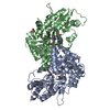
| ||||||||
|---|---|---|---|---|---|---|---|---|---|
| 1 |
| ||||||||
| Unit cell |
|
-Components
| #1: Protein | Mass: 46224.977 Da / Num. of mol.: 2 Source method: isolated from a genetically manipulated source Source: (gene. exp.)   Spodoptera frugiperda (fall armyworm) Spodoptera frugiperda (fall armyworm)References: UniProt: Q71RI9, kynurenine-oxoglutarate transaminase, cysteine-S-conjugate beta-lyase #2: Chemical |   Type: L-peptide linking / Mass: 146.144 Da / Num. of mol.: 2 / Source method: obtained synthetically / Formula: C5H10N2O3 Type: L-peptide linking / Mass: 146.144 Da / Num. of mol.: 2 / Source method: obtained synthetically / Formula: C5H10N2O3#3: Chemical |   Mass: 248.173 Da / Num. of mol.: 2 / Source method: obtained synthetically / Formula: C8H13N2O5P Mass: 248.173 Da / Num. of mol.: 2 / Source method: obtained synthetically / Formula: C8H13N2O5P#4: Chemical | ChemComp-GOL /   Mass: 92.094 Da / Num. of mol.: 4 / Source method: obtained synthetically / Formula: C3H8O3 Mass: 92.094 Da / Num. of mol.: 4 / Source method: obtained synthetically / Formula: C3H8O3#5: Water | ChemComp-HOH / |  Mass: 18.015 Da / Num. of mol.: 411 / Source method: isolated from a natural source / Formula: H2O Mass: 18.015 Da / Num. of mol.: 411 / Source method: isolated from a natural source / Formula: H2O |
|---|
-Experimental details
-Experiment
| Experiment | Method: X-RAY DIFFRACTION / Number of used crystals: 1 |
|---|
- Sample preparation
| Crystal | Density Matthews: 2.38 Å3/Da / Density % sol: 48.33 % |
|---|---|
| Crystal grow | Temperature: 277 K / Method: vapor diffusion, hanging drop / pH: 7.5 Details: 21% PEG 400, 150 mM CaCl2, 10% glycerol, 100 mM HEPES, pH 7.5, VAPOR DIFFUSION, HANGING DROP, temperature 277K |
-Data collection
| Diffraction | Mean temperature: 100 K |
|---|---|
| Diffraction source | Source: SYNCHROTRON / Site: NSLS  / Beamline: X29A / Wavelength: 1.0809 Å / Beamline: X29A / Wavelength: 1.0809 Å |
| Detector | Type: ADSC QUANTUM 315 / Detector: CCD / Date: Jun 27, 2008 |
| Radiation | Protocol: SINGLE WAVELENGTH / Monochromatic (M) / Laue (L): M / Scattering type: x-ray |
| Radiation wavelength | Wavelength: 1.0809 Å / Relative weight: 1 |
| Reflection | Resolution: 2.26→30 Å / Num. all: 47345 / Num. obs: 45176 / % possible obs: 95.4 % / Redundancy: 10.3 % / Rmerge(I) obs: 0.09 |
| Reflection shell | Resolution: 2.26→2.34 Å / Redundancy: 2.1 % / Rmerge(I) obs: 0.43 |
- Processing
| Software |
| ||||||||||||||||||||||||||||||||||||||||||||||||||||||||||||||||||||||||||||||||||||||||||
|---|---|---|---|---|---|---|---|---|---|---|---|---|---|---|---|---|---|---|---|---|---|---|---|---|---|---|---|---|---|---|---|---|---|---|---|---|---|---|---|---|---|---|---|---|---|---|---|---|---|---|---|---|---|---|---|---|---|---|---|---|---|---|---|---|---|---|---|---|---|---|---|---|---|---|---|---|---|---|---|---|---|---|---|---|---|---|---|---|---|---|---|
| Refinement | Method to determine structure: MOLECULAR REPLACEMENT Starting model: PDB entry 2ZJG Resolution: 2.26→29.87 Å / Cor.coef. Fo:Fc: 0.952 / Cor.coef. Fo:Fc free: 0.921 / Cross valid method: THROUGHOUT / ESU R: 0.281 / ESU R Free: 0.209 / Stereochemistry target values: MAXIMUM LIKELIHOOD
| ||||||||||||||||||||||||||||||||||||||||||||||||||||||||||||||||||||||||||||||||||||||||||
| Solvent computation | Ion probe radii: 0.8 Å / Shrinkage radii: 0.8 Å / VDW probe radii: 1.2 Å / Solvent model: MASK | ||||||||||||||||||||||||||||||||||||||||||||||||||||||||||||||||||||||||||||||||||||||||||
| Displacement parameters | Biso mean: 27.831 Å2
| ||||||||||||||||||||||||||||||||||||||||||||||||||||||||||||||||||||||||||||||||||||||||||
| Refinement step | Cycle: LAST / Resolution: 2.26→29.87 Å
| ||||||||||||||||||||||||||||||||||||||||||||||||||||||||||||||||||||||||||||||||||||||||||
| Refine LS restraints |
| ||||||||||||||||||||||||||||||||||||||||||||||||||||||||||||||||||||||||||||||||||||||||||
| LS refinement shell | Resolution: 2.26→2.32 Å / Total num. of bins used: 20
|
