 Movie
Movie Controller

[English] 日本語
 Yorodumi
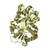
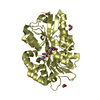
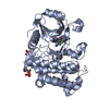
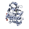
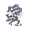
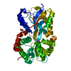
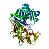
- PDB-3e13: Iron reconstituted ferric binding protein from Campylobacter jejuni -
+ Open data

- Basic information
| Entry | Database: PDB / ID: 30000000000000 | ||||||
|---|---|---|---|---|---|---|---|
| Title | Iron reconstituted ferric binding protein from Campylobacter jejuni | ||||||
 Components Components | Putative iron-uptake ABC transport system,periplasmic iron-binding protein | ||||||
 Keywords Keywords | METAL BINDING PROTEIN / Beta sheet surrounded by alpha helices / anti-parallel beta sheet containing hinge region | ||||||
| Function / homology |  Function and homology information Function and homology information | ||||||
| Biological species |   Campylobacter jejuni (Campylobacter) Campylobacter jejuni (Campylobacter) | ||||||
| Method |  X-RAY DIFFRACTION / SYNCHROTRON / MOLECULAR REPLACEMENT / Resolution: 1.6 Å X-RAY DIFFRACTION / SYNCHROTRON / MOLECULAR REPLACEMENT / Resolution: 1.6 Å | ||||||
 Authors Authors | Murphy, M.E.P. / Tom-Yew, S.A.L. / Bekker, E.G. | ||||||
 Citation Citation | Journal: To be Published Title: Small hinge motion by the anion-independent ferric binding protein from Campylobacter jejuni Authors: Tom-Yew, S.A.L. / Shilton, B.H. / Bekker, E.G. / Gaynor, E.C. / Murphy, M.E.P. | ||||||
| History |
|
- Structure visualization
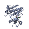
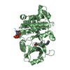
| Structure viewer | Molecule: MolmilJmol/JSmol |
|---|
- Downloads & links
-Download
| PDBx/mmCIF format | 3e13.cif.gz | 87.8 KB | Display | PDBx/mmCIF format |
|---|---|---|---|---|
| PDB format | pdb3e13.ent.gz | 64.3 KB | Display | PDB format |
| PDBx/mmJSON format | 3e13.json.gz | Tree view | PDBx/mmJSON format | |
| Others |  Other downloads Other downloads |
-Validation report
| Arichive directory | https://data.pdbj.org/pub/pdb/validation_reports/e1/3e13ftp://data.pdbj.org/pub/pdb/validation_reports/e1/3e13 | HTTPS FTP |
|---|
-Related structure data
| Related structure data | 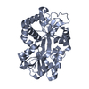 1y4tS S: Starting model for refinement |
|---|---|
| Similar structure data |

-Links
 PDBj
PDBj


- Assembly
| Deposited unit | 
| ||||||||
|---|---|---|---|---|---|---|---|---|---|
| 1 |
| ||||||||
| Unit cell |
|
-Components
| #1: Protein | Mass: 36119.992 Da / Num. of mol.: 1 Source method: isolated from a genetically manipulated source Source: (gene. exp.) Campylobacter jejuni (Campylobacter) / Strain: NCTC 11168 / Gene: cfbpA, Cj0175c / Plasmid: pET28a / Production host: |
|---|---|
| #2: Chemical | ChemComp-FE /   Mass: 55.845 Da / Num. of mol.: 1 / Source method: obtained synthetically / Formula: Fe Mass: 55.845 Da / Num. of mol.: 1 / Source method: obtained synthetically / Formula: Fe |
| #3: Water | ChemComp-HOH /  Mass: 18.015 Da / Num. of mol.: 480 / Source method: isolated from a natural source / Formula: H2O Mass: 18.015 Da / Num. of mol.: 480 / Source method: isolated from a natural source / Formula: H2O |
-Experimental details
-Experiment
| Experiment | Method: X-RAY DIFFRACTION / Number of used crystals: 1 |
|---|
- Sample preparation
| Crystal | Density Matthews: 2 Å3/Da / Density % sol: 38.37 % |
|---|---|
| Crystal grow | Temperature: 298 K / Method: vapor diffusion, hanging drop / pH: 8.5 Details: 24% PEG 4000, 0.1M Tris-HCl, 60 mM sodium acetate, pH 8.5, VAPOR DIFFUSION, HANGING DROP, temperature 298K |
-Data collection
| Diffraction | Mean temperature: 100 K |
|---|---|
| Diffraction source | Source: SYNCHROTRON / Site: SSRL  / Beamline: BL11-3 / Wavelength: 0.974609 Å / Beamline: BL11-3 / Wavelength: 0.974609 Å |
| Detector | Type: ADSC QUANTUM 4 / Detector: CCD / Date: Jun 20, 2004 Details: Flat mirror (vertical focusing) ; single crystal (311) bent monochromator (horizontal focusing) |
| Radiation | Monochromator: Si(311) / Protocol: SINGLE WAVELENGTH / Monochromatic (M) / Laue (L): M / Scattering type: x-ray |
| Radiation wavelength | Wavelength: 0.974609 Å / Relative weight: 1 |
| Reflection | Resolution: 1.6→48.8 Å / Num. obs: 38962 / % possible obs: 90.9 % / Observed criterion σ(F): 0 / Observed criterion σ(I): 0 / Redundancy: 12.9 % / Biso Wilson estimate: 13.3 Å2 / Rmerge(I) obs: 0.106 / Net I/σ(I): 8.6 |
| Reflection shell | Resolution: 1.6→1.66 Å / Rmerge(I) obs: 0.196 / Mean I/σ(I) obs: 3 / % possible all: 73.9 |
- Processing
| Software |
| ||||||||||||||||||||||||||||||||||||||||||||||||||||||||||||||||||||||||||||||||||||||||||||||||||||
|---|---|---|---|---|---|---|---|---|---|---|---|---|---|---|---|---|---|---|---|---|---|---|---|---|---|---|---|---|---|---|---|---|---|---|---|---|---|---|---|---|---|---|---|---|---|---|---|---|---|---|---|---|---|---|---|---|---|---|---|---|---|---|---|---|---|---|---|---|---|---|---|---|---|---|---|---|---|---|---|---|---|---|---|---|---|---|---|---|---|---|---|---|---|---|---|---|---|---|---|---|---|
| Refinement | Method to determine structure: MOLECULAR REPLACEMENT Starting model: PDB entry 1Y4T Resolution: 1.6→48.8 Å / Cor.coef. Fo:Fc: 0.968 / Cor.coef. Fo:Fc free: 0.949 / SU B: 1.552 / SU ML: 0.054 / Cross valid method: THROUGHOUT / σ(F): 0 / ESU R: 0.09 / ESU R Free: 0.093 / Stereochemistry target values: MAXIMUM LIKELIHOOD / Details: HYDROGENS HAVE BEEN ADDED IN THE RIDING POSITIONS
| ||||||||||||||||||||||||||||||||||||||||||||||||||||||||||||||||||||||||||||||||||||||||||||||||||||
| Solvent computation | Ion probe radii: 0.8 Å / Shrinkage radii: 0.8 Å / VDW probe radii: 1.4 Å / Solvent model: BABINET MODEL WITH MASK | ||||||||||||||||||||||||||||||||||||||||||||||||||||||||||||||||||||||||||||||||||||||||||||||||||||
| Displacement parameters | Biso mean: 11.729 Å2
| ||||||||||||||||||||||||||||||||||||||||||||||||||||||||||||||||||||||||||||||||||||||||||||||||||||
| Refinement step | Cycle: LAST / Resolution: 1.6→48.8 Å
| ||||||||||||||||||||||||||||||||||||||||||||||||||||||||||||||||||||||||||||||||||||||||||||||||||||
| Refine LS restraints |
| ||||||||||||||||||||||||||||||||||||||||||||||||||||||||||||||||||||||||||||||||||||||||||||||||||||
| LS refinement shell | Resolution: 1.6→1.642 Å / Total num. of bins used: 20 /
|
