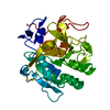
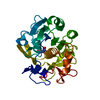
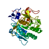
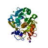
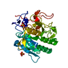
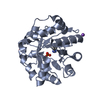
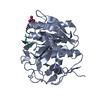
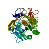
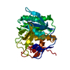
ProteinaseK / Tritirachium alkaline proteinase / Endopeptidase K
Mass: 28958.791 Da / Num. of mol.: 1 / Source method: isolated from a natural source / Source: (natural) Engyodontium album (fungus) / References: UniProt: P06873, peptidase K
Mass: 18.015 Da / Num. of mol.: 341 / Source method: isolated from a natural source / Formula: H2O
Has protein modification
Y
Sequence details
AUTHORS STATE THAT ACCORDING TO THE ELECTRON DENSITY MAPS, RESIDUE 207 SHOULD BE AN ASP. THIS IS ...AUTHORS STATE THAT ACCORDING TO THE ELECTRON DENSITY MAPS, RESIDUE 207 SHOULD BE AN ASP. THIS IS CONSISTENT WITH THE OBSERVATIONS FOUND IN PDB ENTRIES 1IC6, 1P7V AND 2PWA.
-
Experimental details
-
Experiment
Experiment
Method: X-RAY DIFFRACTION / Number of used crystals: 1
-
Sample preparation
Crystal
Density Matthews: 2.02 Å3/Da / Density % sol: 39.02 %
Crystal grow
Temperature: 295 K / Method: vapor diffusion, sitting drop / pH: 7 Details: 5% PEG 3350, 0.1 M HEPES; reservoir of 25% PEG 3350, pH 7.0, VAPOR DIFFUSION, SITTING DROP, temperature 295K
Resolution: 1.32→30.36 Å / Cor.coef. Fo:Fc: 0.977 / Cor.coef. Fo:Fc free: 0.964 / SU B: 2.839 / SU ML: 0.049 / Isotropic thermal model: ANISOTROPIC / Cross valid method: THROUGHOUT / ESU R: 0.067 / ESU R Free: 0.064 / Stereochemistry target values: MAXIMUM LIKELIHOOD Details: HYDROGENS FOR ALL NON-WATER MOLECULES HAVE BEEN ADDED IN THE RIDING POSITION AND ADJUSTED TO AVOID STERIC PROBLEMS. OCCUPANCIES FOR HYDROGENS WERE SET TO THE OCCUPANCY OF THE ATOM TO WHICH ...Details: HYDROGENS FOR ALL NON-WATER MOLECULES HAVE BEEN ADDED IN THE RIDING POSITION AND ADJUSTED TO AVOID STERIC PROBLEMS. OCCUPANCIES FOR HYDROGENS WERE SET TO THE OCCUPANCY OF THE ATOM TO WHICH THE HYDROGEN WAS ATTACHED. THE ATOMS OF AD0 HAVING ZERO OCCUPANCY WERE INCLUDED AS A COMPLETE DESCRIPTION OF THE LIGAND. THERE WAS NO ELECTRON DENSITY TO INDICATE WHERE THIS PYRANOSE RING BELONGED. HOWEVER, THERE WAS DENSITY SUGGESTIVE OF WATER MOLECULES IN THIS REGION AND, THEREFORE, WAS MODELED AS SUCH. HENCE, THERE APPEARS TO BE CLOSE CONTACTS AS SHOWN IN REMARK 500. THERE IS EXCELLENT DENSITY FOR EACH OF THE RESIDUES DESCRIBED IN REMARK 500 AS RAMACHANDRAN OUTLIERS.
Rfactor
Num. reflection
% reflection
Selection details
Rfree
0.20669
2646
5.1 %
RANDOM
Rwork
0.1579
-
-
-
all
0.16036
49454
-
-
obs
0.16036
49454
92.33 %
-
Solvent computation
Ion probe radii: 0.8 Å / Shrinkage radii: 0.8 Å / VDW probe radii: 1.4 Å / Solvent model: BABINET MODEL WITH MASK
Displacement parameters
Biso mean: 15.875 Å2
Baniso -1
Baniso -2
Baniso -3
1-
-0.45 Å2
0 Å2
0 Å2
2-
-
-0.45 Å2
0 Å2
3-
-
-
0.9 Å2
Refinement step
Cycle: LAST / Resolution: 1.32→30.36 Å
Protein
Nucleic acid
Ligand
Solvent
Total
Num. atoms
2032
0
42
341
2415
Refine LS restraints
Refine-ID
Type
Dev ideal
Dev ideal target
Number
X-RAY DIFFRACTION
r_bond_refined_d
0.011
0.021
2199
X-RAY DIFFRACTION
r_bond_other_d
0
0.02
2067
X-RAY DIFFRACTION
r_angle_refined_deg
1.265
1.951
3015
X-RAY DIFFRACTION
r_angle_other_deg
0.674
3
4665
X-RAY DIFFRACTION
r_dihedral_angle_1_deg
6.234
5
311
X-RAY DIFFRACTION
r_dihedral_angle_2_deg
35.854
23.736
91
X-RAY DIFFRACTION
r_dihedral_angle_3_deg
12.819
15
329
X-RAY DIFFRACTION
r_dihedral_angle_4_deg
19.321
15
14
X-RAY DIFFRACTION
r_chiral_restr
0.078
0.2
335
X-RAY DIFFRACTION
r_gen_planes_refined
0.01
0.02
2566
X-RAY DIFFRACTION
r_gen_planes_other
0
0.02
537
X-RAY DIFFRACTION
r_mcbond_it
2.568
2
1422
X-RAY DIFFRACTION
r_mcbond_other
2.262
2
601
X-RAY DIFFRACTION
r_mcangle_it
3.353
4
2281
X-RAY DIFFRACTION
r_scbond_it
4.085
4
777
X-RAY DIFFRACTION
r_scangle_it
5.315
6
717
X-RAY DIFFRACTION
r_rigid_bond_restr
2.377
3
2198
X-RAY DIFFRACTION
r_sphericity_free
8.481
3
380
X-RAY DIFFRACTION
r_sphericity_bonded
4.871
3
2140
LS refinement shell
Refine-ID: X-RAY DIFFRACTION / Total num. of bins used: 20
Resolution (Å)
Rfactor Rfree
Num. reflection Rfree
Rfactor Rwork
Num. reflection Rwork
Rfactor all
Num. reflection obs
% reflection obs (%)
1.319-1.353
0.475
67
0.456
1119
0.457
4114
28.83
1.353-1.39
0.474
163
0.394
2876
0.399
3990
76.16
1.39-1.431
0.425
185
0.376
3445
0.379
3874
93.7
1.431-1.475
0.4
198
0.331
3589
0.334
3812
99.34
1.475-1.523
0.406
185
0.297
3441
0.303
3644
99.51
1.523-1.576
0.307
152
0.255
3394
0.257
3560
99.61
1.576-1.636
0.306
162
0.211
3254
0.216
3429
99.62
1.636-1.702
0.266
170
0.177
3139
0.182
3323
99.58
1.702-1.778
0.222
172
0.148
3002
0.152
3188
99.56
1.778-1.864
0.217
157
0.126
2869
0.131
3044
99.41
1.864-1.964
0.178
137
0.115
2755
0.118
2905
99.55
1.964-2.083
0.176
132
0.113
2622
0.116
2764
99.64
2.083-2.226
0.175
142
0.115
2455
0.118
2611
99.46
2.226-2.403
0.145
141
0.117
2279
0.119
2425
99.79
2.403-2.631
0.169
113
0.118
2124
0.12
2248
99.51
2.631-2.939
0.157
108
0.123
1947
0.125
2062
99.66
2.939-3.388
0.159
97
0.118
1730
0.12
1836
99.51
3.388-4.137
0.145
76
0.112
1482
0.114
1563
99.68
4.137-5.797
0.18
50
0.163
1203
0.164
1257
99.68
5.797-30.359
0.179
39
0.224
729
0.222
776
98.97
+
About Yorodumi
-
News
-
Feb 9, 2022. New format data for meta-information of EMDB entries
New format data for meta-information of EMDB entries
Version 3 of the EMDB header file is now the official format.
The previous official version 1.9 will be removed from the archive.
In the structure databanks used in Yorodumi, some data are registered as the other names, "COVID-19 virus" and "2019-nCoV". Here are the details of the virus and the list of structure data.
Jan 31, 2019. EMDB accession codes are about to change! (news from PDBe EMDB page)
EMDB accession codes are about to change! (news from PDBe EMDB page)
The allocation of 4 digits for EMDB accession codes will soon come to an end. Whilst these codes will remain in use, new EMDB accession codes will include an additional digit and will expand incrementally as the available range of codes is exhausted. The current 4-digit format prefixed with “EMD-” (i.e. EMD-XXXX) will advance to a 5-digit format (i.e. EMD-XXXXX), and so on. It is currently estimated that the 4-digit codes will be depleted around Spring 2019, at which point the 5-digit format will come into force.
The EM Navigator/Yorodumi systems omit the EMD- prefix.
Related info.:Q: What is EMD? / ID/Accession-code notation in Yorodumi/EM Navigator
Yorodumi is a browser for structure data from EMDB, PDB, SASBDB, etc.
This page is also the successor to EM Navigator detail page, and also detail information page/front-end page for Omokage search.
The word "yorodu" (or yorozu) is an old Japanese word meaning "ten thousand". "mi" (miru) is to see.
Related info.:EMDB / PDB / SASBDB / Comparison of 3 databanks / Yorodumi Search / Aug 31, 2016. New EM Navigator & Yorodumi / Yorodumi Papers / Jmol/JSmol / Function and homology information / Changes in new EM Navigator and Yorodumi
 Movie
Movie Controller


 Open data
 Basic information
 Components
 Keywords
 Function and homology information
 Engyodontium album (fungus)
 X-RAY DIFFRACTION /
  Authors
 Citation
 Structure visualization
 Downloads & links
 Other downloads
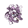

 PDBj
PDBj

 Assembly
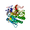

 Mass: 40.078 Da / Num. of mol.: 2 / Source method: obtained synthetically / Formula: Ca
 Mass: 238.305 Da / Num. of mol.: 1 / Source method: obtained synthetically / Formula: C8H18N2O4S / Comment: pH buffer*YM
 Mass: 18.015 Da / Num. of mol.: 341 / Source method: isolated from a natural source / Formula: H2O
 Sample preparation
 Processing