 Movie
Movie Controller

[English] 日本語
 Yorodumi
- PDB-3dto: Crystal structure of the metal-dependent HD domain-containing hyd... -
+ Open data

- Basic information
| Entry | Database: PDB / ID: 3dto | ||||||
|---|---|---|---|---|---|---|---|
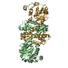
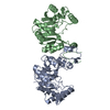
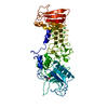
| Title | Crystal structure of the metal-dependent HD domain-containing hydrolase BH2835 from Bacillus halodurans, Northeast Structural Genomics Consortium Target BhR130. | ||||||
 Components Components | BH2835 protein | ||||||
 Keywords Keywords | structural genomics / unknown function / all alpha-helical protein / PSI-2 / Protein Structure Initiative / Northeast Structural Genomics Consortium / NESG | ||||||
| Function / homology |  Function and homology information Function and homology informationHD-domain/PDEase-like / Methane Monooxygenase Hydroxylase; Chain G, domain 1 - #1910 / HD domain / HD domain / Cyclin A; domain 1 / Metal dependent phosphohydrolases with conserved 'HD' motif. / HD/PDEase domain / Methane Monooxygenase Hydroxylase; Chain G, domain 1 / Up-down Bundle / Orthogonal Bundle / Mainly Alpha Similarity search - Domain/homology | ||||||
| Biological species |  Bacillus halodurans (bacteria) Bacillus halodurans (bacteria) | ||||||
| Method |  X-RAY DIFFRACTION / SYNCHROTRON / SAD / Resolution: 3.3 Å X-RAY DIFFRACTION / SYNCHROTRON / SAD / Resolution: 3.3 Å | ||||||
 Authors Authors | Forouhar, F. / Su, M. / Seetharaman, J. / Janjua, H. / Fang, Y. / Xiao, R. / Cunningham, K. / Ma, L.-C. / Owen, L.A. / Wang, D. ...Forouhar, F. / Su, M. / Seetharaman, J. / Janjua, H. / Fang, Y. / Xiao, R. / Cunningham, K. / Ma, L.-C. / Owen, L.A. / Wang, D. / Tong, S. / Everett, J.K. / Acton, T.B. / Montelione, G.T. / Tong, L. / Hunt, J.F. / Northeast Structural Genomics Consortium (NESG) | ||||||
 Citation Citation | Journal: To be Published Title: Crystal structure of the metal-dependent HD domain-containing hydrolase BH2835 from Bacillus halodurans, Northeast Structural Genomics Consortium Target BhR130. Authors: Forouhar, F. / Su, M. / Seetharaman, J. / Janjua, H. / Fang, Y. / Xiao, R. / Cunningham, K. / Ma, L.-C. / Owen, L.A. / Wang, D. / Tong, S. / Everett, J.K. / Acton, T.B. / Montelione, G.T. / Tong, L. / Hunt, J.F. | ||||||
| History |
|
- Structure visualization
| Structure viewer | Molecule: MolmilJmol/JSmol |
|---|
- Downloads & links
-Download
| PDBx/mmCIF format | 3dto.cif.gz | 160.9 KB | Display | PDBx/mmCIF format |
|---|---|---|---|---|
| PDB format | pdb3dto.ent.gz | 130.3 KB | Display | PDB format |
| PDBx/mmJSON format | 3dto.json.gz | Tree view | PDBx/mmJSON format | |
| Others |  Other downloads Other downloads |
-Validation report
| Arichive directory | https://data.pdbj.org/pub/pdb/validation_reports/dt/3dtoftp://data.pdbj.org/pub/pdb/validation_reports/dt/3dto | HTTPS FTP |
|---|
-Related structure data
| Similar structure data | |
|---|---|
| Other databases |


-Links
 PDBj
PDBj
- Assembly
| Deposited unit | 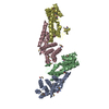
| ||||||||
|---|---|---|---|---|---|---|---|---|---|
| 1 | 
| ||||||||
| 2 | 
| ||||||||
| Unit cell |
|
-Components
| #1: Protein | Mass: 26204.098 Da / Num. of mol.: 4 Source method: isolated from a genetically manipulated source Source: (gene. exp.) Bacillus halodurans (bacteria) / Strain: C-125 / Gene: BH2835 / Plasmid: pET21 / Production host: Has protein modification | Y | |
|---|
-Experimental details
-Experiment
| Experiment | Method: X-RAY DIFFRACTION / Number of used crystals: 1 |
|---|
- Sample preparation
| Crystal | Density Matthews: 2.48 Å3/Da / Density % sol: 50.45 % |
|---|---|
| Crystal grow | Temperature: 291 K / Method: microbatch under oil / pH: 7 Details: Protein solution: 10 mM Tris (pH 7.5), 100 mM sodium chloride, and 5 mM DTT. Reservoir solution: 0.1M mops (pH 7), 80% PEG 400, and 0.1M potassium acetate. , microbatch under oil, temperature 291K |
-Data collection
| Diffraction | Mean temperature: 100 K |
|---|---|
| Diffraction source | Source: SYNCHROTRON / Site: NSLS  / Beamline: X4A / Wavelength: 0.97915 Å / Beamline: X4A / Wavelength: 0.97915 Å |
| Detector | Type: ADSC QUANTUM 4 / Detector: CCD / Date: Aug 19, 2007 / Details: mirrors. |
| Radiation | Monochromator: Si 111 CHANNEL / Protocol: SINGLE WAVELENGTH / Monochromatic (M) / Laue (L): M / Scattering type: x-ray |
| Radiation wavelength | Wavelength: 0.97915 Å / Relative weight: 1 |
| Reflection | Resolution: 3.3→30 Å / Num. obs: 22713 / % possible obs: 74.5 % / Observed criterion σ(F): 0.5 / Observed criterion σ(I): 0.5 / Redundancy: 3.4 % / Biso Wilson estimate: 74.8 Å2 / Rmerge(I) obs: 0.081 / Rsym value: 0.075 / Net I/σ(I): 13.1 |
| Reflection shell | Resolution: 3.3→3.42 Å / Redundancy: 1.9 % / Rmerge(I) obs: 0.149 / Mean I/σ(I) obs: 3 / Rsym value: 0.185 / % possible all: 58.4 |
- Processing
| Software |
| ||||||||||||||||||||||||||||||||||||||||||||||||||||||||||||
|---|---|---|---|---|---|---|---|---|---|---|---|---|---|---|---|---|---|---|---|---|---|---|---|---|---|---|---|---|---|---|---|---|---|---|---|---|---|---|---|---|---|---|---|---|---|---|---|---|---|---|---|---|---|---|---|---|---|---|---|---|---|
| Refinement | Method to determine structure: SAD / Resolution: 3.3→19.76 Å / Rfactor Rfree error: 0.006 / Data cutoff high absF: 335033.32 / Data cutoff low absF: 0 / Isotropic thermal model: RESTRAINED / Cross valid method: THROUGHOUT / σ(F): 2 / Stereochemistry target values: Engh & Huber
| ||||||||||||||||||||||||||||||||||||||||||||||||||||||||||||
| Solvent computation | Solvent model: FLAT MODEL / Bsol: 45.1144 Å2 / ksol: 0.35 e/Å3 | ||||||||||||||||||||||||||||||||||||||||||||||||||||||||||||
| Displacement parameters | Biso mean: 70.3 Å2
| ||||||||||||||||||||||||||||||||||||||||||||||||||||||||||||
| Refine analyze |
| ||||||||||||||||||||||||||||||||||||||||||||||||||||||||||||
| Refinement step | Cycle: LAST / Resolution: 3.3→19.76 Å
| ||||||||||||||||||||||||||||||||||||||||||||||||||||||||||||
| Refine LS restraints |
| ||||||||||||||||||||||||||||||||||||||||||||||||||||||||||||
| LS refinement shell | Resolution: 3.3→3.42 Å / Rfactor Rfree error: 0.027 / Total num. of bins used: 10
|
