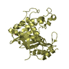
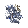
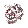
- PDB-3dka: Crystal structure of a dinb-like protein (yjoa, bsu12410) from ba... -
+
Open data
ID or keywords:
Loading...
-
Basic information
Entry
Database: PDB / ID: 3dka
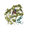
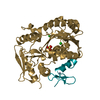
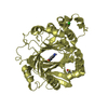
Title
Crystal structure of a dinb-like protein (yjoa, bsu12410) from bacillus subtilis at 2.30 A resolution
Components
DinB-like Protein
Keywords
UNKNOWN FUNCTION / Dinb/yfit-like putative metalloenzyme fold / structural genomics / Joint Center for Structural Genomics / JCSG / Protein Structure Initiative / PSI-2
Function / homology
DNA damage-inducible protein DinB / DinB family / dinb family like domain / DinB/YfiT-like putative metalloenzymes / Four Helix Bundle (Hemerythrin (Met), subunit A) / Up-down Bundle / Mainly Alpha / metal ion binding / Uncharacterized protein YjoA
Function and homology information
Biological species
Bacillus subtilis (bacteria)
Method
X-RAY DIFFRACTION / SYNCHROTRON / MAD / Resolution: 2.3 Å
Mass: 18.015 Da / Num. of mol.: 98 / Source method: isolated from a natural source / Formula: H2O
Has protein modification
Y
Sequence details
THE CONSTRUCT WAS EXPRESSED WITH A PURIFICATION TAG MGSDKIHHHHHHENLYFQG. THE TAG WAS REMOVED WITH ...THE CONSTRUCT WAS EXPRESSED WITH A PURIFICATION TAG MGSDKIHHHHHHENLYFQG. THE TAG WAS REMOVED WITH TEV PROTEASE LEAVING ONLY A GLYCINE (0) FOLLOWED BY THE TARGET SEQUENCE.
-
Experimental details
-
Experiment
Experiment
Method: X-RAY DIFFRACTION / Number of used crystals: 1
-
Sample preparation
Crystal
Density Matthews: 2.07 Å3/Da / Density % sol: 40.57 %
Crystal grow
Temperature: 277 K / Method: vapor diffusion, sitting drop / pH: 5.6 Details: 0.2000M NH4OAc, 30.0000% PEG-4000, 0.1M Citrate pH 5.6, NANODROP, VAPOR DIFFUSION, SITTING DROP, temperature 277K
Type: MARMOSAIC 325 mm CCD / Detector: CCD / Date: May 14, 2008 / Details: Flat mirror (vertical focusing)
Radiation
Monochromator: Single crystal Si(111) bent monochromator (horizontal focusing) Protocol: MAD / Monochromatic (M) / Laue (L): M / Scattering type: x-ray
Radiation wavelength
ID
Wavelength (Å)
Relative weight
1
0.91837
1
2
0.97956
1
3
0.97904
1
Reflection
Resolution: 2.3→29.566 Å / Num. obs: 13596 / % possible obs: 95.9 % / Observed criterion σ(I): -3 / Redundancy: 3.63 % / Biso Wilson estimate: 34.972 Å2 / Rmerge(I) obs: 0.097 / Net I/σ(I): 7.3
Reflection shell
Resolution (Å)
Rmerge(I) obs
Mean I/σ(I) obs
Num. measured obs
Num. unique obs
Diffraction-ID
% possible all
2.3-2.38
0.571
1.58
4548
2313
1
90.5
2.38-2.48
0.462
1.9
5203
2619
1
96.1
2.48-2.59
0.378
2.4
4812
2441
1
96.4
2.59-2.73
0.28
3
5069
2554
1
96
2.73-2.9
0.214
3.9
4885
2460
1
96
2.9-3.12
0.163
5
4873
2468
1
97.2
3.12-3.43
0.107
7.6
4973
2501
1
96.8
3.43-3.93
0.06
12
4994
2523
1
96.4
3.93-4.93
0.04
16.6
4955
2499
1
97.1
4.93-29.57
0.035
18.5
5051
2562
1
96.5
-
Phasing
Phasing
Method: MAD
-
Processing
Software
Name
Version
Classification
NB
REFMAC
5.2.0019
refinement
PHENIX
refinement
SHELX
phasing
MolProbity
3beta29
modelbuilding
XSCALE
datascaling
PDB_EXTRACT
3.004
dataextraction
XDS
datareduction
SHELXD
phasing
autoSHARP
phasing
Refinement
Method to determine structure: MAD / Resolution: 2.3→29.566 Å / Cor.coef. Fo:Fc: 0.93 / Cor.coef. Fo:Fc free: 0.893 / SU B: 15.654 / SU ML: 0.193 / TLS residual ADP flag: LIKELY RESIDUAL / Cross valid method: THROUGHOUT / σ(F): 0 / ESU R: 0.437 / ESU R Free: 0.273 Stereochemistry target values: MAXIMUM LIKELIHOOD WITH PHASES Details: 1. HYDROGENS HAVE BEEN ADDED IN THE RIDING POSITIONS. 2. A MET-INHIBITION PROTOCOL WAS USED FOR SELENOMETHIONINE INCORPORATION DURING PROTEIN EXPRESSION. THE OCCUPANCY OF THE SE ATOMS IN THE ...Details: 1. HYDROGENS HAVE BEEN ADDED IN THE RIDING POSITIONS. 2. A MET-INHIBITION PROTOCOL WAS USED FOR SELENOMETHIONINE INCORPORATION DURING PROTEIN EXPRESSION. THE OCCUPANCY OF THE SE ATOMS IN THE MSE RESIDUES WAS REDUCED TO 0.75 FOR THE REDUCED SCATTERING POWER DUE TO PARTIAL S-MET INCORPORATION. 3. ATOM RECORDS CONTAIN RESIDUAL B FACTORS ONLY.
Rfactor
Num. reflection
% reflection
Selection details
Rfree
0.269
677
5 %
RANDOM
Rwork
0.217
-
-
-
obs
0.219
13568
97.56 %
-
Solvent computation
Ion probe radii: 0.8 Å / Shrinkage radii: 0.8 Å / VDW probe radii: 1.2 Å / Solvent model: MASK
In the structure databanks used in Yorodumi, some data are registered as the other names, "COVID-19 virus" and "2019-nCoV". Here are the details of the virus and the list of structure data.
Jan 31, 2019. EMDB accession codes are about to change! (news from PDBe EMDB page)
EMDB accession codes are about to change! (news from PDBe EMDB page)
The allocation of 4 digits for EMDB accession codes will soon come to an end. Whilst these codes will remain in use, new EMDB accession codes will include an additional digit and will expand incrementally as the available range of codes is exhausted. The current 4-digit format prefixed with “EMD-” (i.e. EMD-XXXX) will advance to a 5-digit format (i.e. EMD-XXXXX), and so on. It is currently estimated that the 4-digit codes will be depleted around Spring 2019, at which point the 5-digit format will come into force.
The EM Navigator/Yorodumi systems omit the EMD- prefix.
Related info.:Q: What is EMD? / ID/Accession-code notation in Yorodumi/EM Navigator
Yorodumi is a browser for structure data from EMDB, PDB, SASBDB, etc.
This page is also the successor to EM Navigator detail page, and also detail information page/front-end page for Omokage search.
The word "yorodu" (or yorozu) is an old Japanese word meaning "ten thousand". "mi" (miru) is to see.
Related info.:EMDB / PDB / SASBDB / Comparison of 3 databanks / Yorodumi Search / Aug 31, 2016. New EM Navigator & Yorodumi / Yorodumi Papers / Jmol/JSmol / Function and homology information / Changes in new EM Navigator and Yorodumi
 Movie
Movie Controller

 Yorodumi
 Open data
 Basic information
 Components
 Keywords
 Function and homology information
 X-RAY DIFFRACTION /
  Authors
 Citation
 Structure visualization
 Downloads & links
 Other downloads
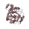


 PDBj
PDBj Assembly
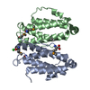

 Mass: 35.453 Da / Num. of mol.: 1 / Source method: obtained synthetically / Formula: Cl
 Mass: 18.015 Da / Num. of mol.: 98 / Source method: isolated from a natural source / Formula: H2O
 Sample preparation
 / Beamline: BL11-1 / Wavelength: 0.91837,0.97956,0.97904
 Processing