#95 - Nov 2007 Multidrug Resistance Transporters similarity (5)
-
Assembly
Deposited unit
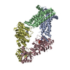
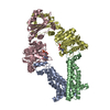
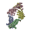
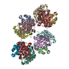
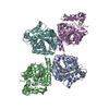
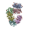
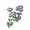
A: D-methionine transport system permease protein metI B: D-methionine transport system permease protein metI C: Methionine import ATP-binding protein metN D: Methionine import ATP-binding protein metN E: D-methionine transport system permease protein metI F: D-methionine transport system permease protein metI G: Methionine import ATP-binding protein metN H: Methionine import ATP-binding protein metN
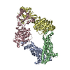
A: D-methionine transport system permease protein metI B: D-methionine transport system permease protein metI C: Methionine import ATP-binding protein metN D: Methionine import ATP-binding protein metN
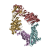
E: D-methionine transport system permease protein metI F: D-methionine transport system permease protein metI G: Methionine import ATP-binding protein metN H: Methionine import ATP-binding protein metN
Authors state that there are two full transporters per asymmetric unit corresponding to chains A,B,C,D and E,F,G,H, respectively.
-
Components
#1: Protein
D-methioninetransportsystempermeaseproteinmetI
Mass: 23269.947 Da / Num. of mol.: 4 Source method: isolated from a genetically manipulated source Source: (gene. exp.) Escherichia coli (E. coli) / Strain: K12 / Gene: metI, yaeE, b0198, JW0194 / Production host: Escherichia coli (E. coli) / References: UniProt: P31547
#2: Protein
MethionineimportATP-bindingproteinmetN
Mass: 37831.281 Da / Num. of mol.: 4 Source method: isolated from a genetically manipulated source Source: (gene. exp.) Escherichia coli (E. coli) / Strain: K12 / Gene: metN, abc, b0199, JW0195 / Production host: Escherichia coli (E. coli) References: UniProt: P30750, Hydrolases; Acting on acid anhydrides; Acting on acid anhydrides to catalyse transmembrane movement of substances
-
Experimental details
-
Experiment
Experiment
Method: X-RAY DIFFRACTION / Number of used crystals: 2
-
Sample preparation
Crystal
Density Matthews: 4.78 Å3/Da / Density % sol: 74.25 %
-
Data collection
Diffraction
ID
Mean temperature (K)
Crystal-ID
1
100
1
2
1
1,2
1
Diffraction source
Source
Site
Beamline
ID
Wavelength (Å)
SYNCHROTRON
SSRL
BL11-1
1
1
SYNCHROTRON
SSRL
BL9-2
2
1.5
Detector
Type
ID
Detector
MARMOSAIC 325 mm CCD
1
CCD
MARMOSAIC 325 mm CCD
2
CCD
Radiation
ID
Protocol
Monochromatic (M) / Laue (L)
Scattering type
Wavelength-ID
1
SINGLEWAVELENGTH
M
x-ray
1
2
SINGLEWAVELENGTH
M
x-ray
2
Radiation wavelength
ID
Wavelength (Å)
Relative weight
1
1
1
2
1.5
1
Reflection
Resolution: 3.7→39.9 Å / Num. obs: 50639
-
Processing
Software
Name
Version
Classification
CNS
1.2
refinement
Blu-Ice
datacollection
MOSFLM
datareduction
SCALA
datascaling
SHARP
phasing
Refinement
Method to determine structure: SAD / Resolution: 3.7→39.9 Å / Rfactor Rfree error: 0.005 / Data cutoff high absF: 16743703.72 / Data cutoff low absF: 0 / Isotropic thermal model: RESTRAINED / Cross valid method: THROUGHOUT / σ(F): 0 / Stereochemistry target values: Engh & Huber / Details: BULK SOLVENT MODEL USED
In the structure databanks used in Yorodumi, some data are registered as the other names, "COVID-19 virus" and "2019-nCoV". Here are the details of the virus and the list of structure data.
Jan 31, 2019. EMDB accession codes are about to change! (news from PDBe EMDB page)
EMDB accession codes are about to change! (news from PDBe EMDB page)
The allocation of 4 digits for EMDB accession codes will soon come to an end. Whilst these codes will remain in use, new EMDB accession codes will include an additional digit and will expand incrementally as the available range of codes is exhausted. The current 4-digit format prefixed with “EMD-” (i.e. EMD-XXXX) will advance to a 5-digit format (i.e. EMD-XXXXX), and so on. It is currently estimated that the 4-digit codes will be depleted around Spring 2019, at which point the 5-digit format will come into force.
The EM Navigator/Yorodumi systems omit the EMD- prefix.
Related info.:Q: What is EMD? / ID/Accession-code notation in Yorodumi/EM Navigator
Yorodumi is a browser for structure data from EMDB, PDB, SASBDB, etc.
This page is also the successor to EM Navigator detail page, and also detail information page/front-end page for Omokage search.
The word "yorodu" (or yorozu) is an old Japanese word meaning "ten thousand". "mi" (miru) is to see.
Related info.:EMDB / PDB / SASBDB / Comparison of 3 databanks / Yorodumi Search / Aug 31, 2016. New EM Navigator & Yorodumi / Yorodumi Papers / Jmol/JSmol / Function and homology information / Changes in new EM Navigator and Yorodumi
 Movie
Movie Controller


 Open data
 Basic information
 Components
 Keywords
 Function and homology information
 X-RAY DIFFRACTION /
  Authors
 Citation
 Structure visualization
 Downloads & links
 Other downloads
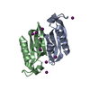

 PDBj
PDBj

 Assembly
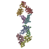
 Sample preparation
 Processing