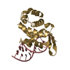
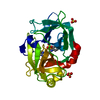
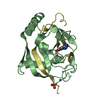
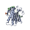
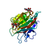
- PDB-3dgt: The 1.5 A crystal structure of endo-1,3-beta-glucanase from Strep... -
+
Open data
ID or keywords:
Loading...
-
Basic information
Entry
Database: PDB / ID: 3dgt
Title
The 1.5 A crystal structure of endo-1,3-beta-glucanase from Streptomyces sioyaensis
Components
Endo-1,3-beta-glucanase
Keywords
HYDROLASE / GHF16 / 1 / 3-beta-glucanase
Function / homology
Function and homology information
hydrolase activity, hydrolyzing O-glycosyl compounds / carbohydrate binding / carbohydrate metabolic process / metal ion binding Similarity search - Function
XgeA-like, catalytic GH16 domain / : / Cellulose binding, type IV / Cellulose Binding Domain Type IV / Glycosyl hydrolases family 16 / Carbohydrate binding module (family 6) / Glycoside hydrolase family 16 / Glycosyl hydrolases family 16 (GH16) domain profile. / CBM6 (carbohydrate binding type-6) domain profile. / Carbohydrate binding module family 6 ...XgeA-like, catalytic GH16 domain / : / Cellulose binding, type IV / Cellulose Binding Domain Type IV / Glycosyl hydrolases family 16 / Carbohydrate binding module (family 6) / Glycoside hydrolase family 16 / Glycosyl hydrolases family 16 (GH16) domain profile. / CBM6 (carbohydrate binding type-6) domain profile. / Carbohydrate binding module family 6 / Jelly Rolls - #200 / Twin arginine translocation (Tat) signal profile. / Twin-arginine translocation pathway, signal sequence / Galactose-binding-like domain superfamily / Concanavalin A-like lectin/glucanase domain superfamily / Jelly Rolls / Sandwich / Mainly Beta Similarity search - Domain/homology
Mass: 18.015 Da / Num. of mol.: 150 / Source method: isolated from a natural source / Formula: H2O
Has protein modification
Y
Sequence details
THERE ARE CONFLICTS BETWEEN THE GIVEN SEQUENCE AND THE DATABASE REFERENCE SEQUENCE. THESE RESIDUES ...THERE ARE CONFLICTS BETWEEN THE GIVEN SEQUENCE AND THE DATABASE REFERENCE SEQUENCE. THESE RESIDUES IDENTIFIED IN THE STRUCTURE DETERMINTION WORK SOULD BE THE CORRECT SEQUENCE OF THE PROTEIN. THE DEPOSITORS ALSO DID THE SEQUENCING OF THE PLASMID AND CONFIRMED THESE RESIDUE SEQUENCES ARE CONSISTENT WITH THE X-RAY STRUCTURE.
-
Experimental details
-
Experiment
Experiment
Method: X-RAY DIFFRACTION / Number of used crystals: 1
-
Sample preparation
Crystal
Density Matthews: 2.03 Å3/Da / Density % sol: 39.54 %
Monochromator: MSC blue-optics / Protocol: SINGLE WAVELENGTH / Monochromatic (M) / Laue (L): M / Scattering type: x-ray
Radiation wavelength
Wavelength: 1.5418 Å / Relative weight: 1
Reflection
Resolution: 1.5→20 Å / Num. obs: 38840 / % possible obs: 99 % / Observed criterion σ(I): -3 / Redundancy: 4.2 % / Biso Wilson estimate: 10.246 Å2 / Rmerge(I) obs: 0.048 / Net I/σ(I): 34.8
Reflection shell
Resolution: 1.5→1.57 Å / Redundancy: 3.39 % / Rmerge(I) obs: 0.098 / Mean I/σ(I) obs: 14.4 / Num. unique all: 4632 / % possible all: 96.3
-
Processing
Software
Name
Version
Classification
REFMAC
5.2.0019
refinement
CrystalClear
datacollection
DENZO
datareduction
SCALEPACK
datascaling
SHELXCD
phasing
SHELXE
modelbuilding
Refinement
Method to determine structure: SAD / Resolution: 1.5→19.75 Å / Cor.coef. Fo:Fc: 0.945 / Cor.coef. Fo:Fc free: 0.939 / SU B: 1.016 / SU ML: 0.04 / Cross valid method: THROUGHOUT / ESU R: 0.079 / ESU R Free: 0.074 / Stereochemistry target values: MAXIMUM LIKELIHOOD / Details: HYDROGENS HAVE BEEN ADDED IN THE RIDING POSITIONS
Rfactor
Num. reflection
% reflection
Selection details
Rfree
0.19832
1949
5 %
RANDOM
Rwork
0.18284
-
-
-
obs
0.18361
36838
99.03 %
-
Solvent computation
Ion probe radii: 0.8 Å / Shrinkage radii: 0.8 Å / VDW probe radii: 1.4 Å / Solvent model: BABINET MODEL WITH MASK
Displacement parameters
Biso mean: 13.559 Å2
Baniso -1
Baniso -2
Baniso -3
1-
-0.01 Å2
0 Å2
0 Å2
2-
-
0 Å2
0 Å2
3-
-
-
0 Å2
Refinement step
Cycle: LAST / Resolution: 1.5→19.75 Å
Protein
Nucleic acid
Ligand
Solvent
Total
Num. atoms
2053
0
1
150
2204
Refine LS restraints
Refine-ID
Type
Dev ideal
Dev ideal target
Number
X-RAY DIFFRACTION
r_bond_refined_d
0.008
0.021
2117
X-RAY DIFFRACTION
r_bond_other_d
X-RAY DIFFRACTION
r_angle_refined_deg
1.244
1.916
2901
X-RAY DIFFRACTION
r_angle_other_deg
X-RAY DIFFRACTION
r_dihedral_angle_1_deg
6.882
5
277
X-RAY DIFFRACTION
r_dihedral_angle_2_deg
38.268
24.624
93
X-RAY DIFFRACTION
r_dihedral_angle_3_deg
10.491
15
274
X-RAY DIFFRACTION
r_dihedral_angle_4_deg
15.983
15
9
X-RAY DIFFRACTION
r_chiral_restr
0.082
0.2
304
X-RAY DIFFRACTION
r_gen_planes_refined
0.004
0.02
1701
X-RAY DIFFRACTION
r_gen_planes_other
X-RAY DIFFRACTION
r_nbd_refined
0.198
0.2
1005
X-RAY DIFFRACTION
r_nbd_other
X-RAY DIFFRACTION
r_nbtor_refined
0.305
0.2
1459
X-RAY DIFFRACTION
r_nbtor_other
X-RAY DIFFRACTION
r_xyhbond_nbd_refined
0.08
0.2
128
X-RAY DIFFRACTION
r_xyhbond_nbd_other
X-RAY DIFFRACTION
r_metal_ion_refined
X-RAY DIFFRACTION
r_metal_ion_other
X-RAY DIFFRACTION
r_symmetry_vdw_refined
0.163
0.2
35
X-RAY DIFFRACTION
r_symmetry_vdw_other
X-RAY DIFFRACTION
r_symmetry_hbond_refined
0.064
0.2
27
X-RAY DIFFRACTION
r_symmetry_hbond_other
X-RAY DIFFRACTION
r_symmetry_metal_ion_refined
X-RAY DIFFRACTION
r_symmetry_metal_ion_other
X-RAY DIFFRACTION
r_mcbond_it
0.476
1.5
1393
X-RAY DIFFRACTION
r_mcbond_other
X-RAY DIFFRACTION
r_mcangle_it
0.811
2
2196
X-RAY DIFFRACTION
r_scbond_it
1.249
3
849
X-RAY DIFFRACTION
r_scangle_it
1.642
4.5
705
X-RAY DIFFRACTION
r_rigid_bond_restr
X-RAY DIFFRACTION
r_sphericity_free
X-RAY DIFFRACTION
r_sphericity_bonded
LS refinement shell
Resolution: 1.5→1.539 Å / Total num. of bins used: 20
Rfactor
Num. reflection
% reflection
Rfree
0.184
153
-
Rwork
0.158
2559
-
obs
-
-
95.19 %
+
About Yorodumi
-
News
-
Feb 9, 2022. New format data for meta-information of EMDB entries
New format data for meta-information of EMDB entries
Version 3 of the EMDB header file is now the official format.
The previous official version 1.9 will be removed from the archive.
In the structure databanks used in Yorodumi, some data are registered as the other names, "COVID-19 virus" and "2019-nCoV". Here are the details of the virus and the list of structure data.
Jan 31, 2019. EMDB accession codes are about to change! (news from PDBe EMDB page)
EMDB accession codes are about to change! (news from PDBe EMDB page)
The allocation of 4 digits for EMDB accession codes will soon come to an end. Whilst these codes will remain in use, new EMDB accession codes will include an additional digit and will expand incrementally as the available range of codes is exhausted. The current 4-digit format prefixed with “EMD-” (i.e. EMD-XXXX) will advance to a 5-digit format (i.e. EMD-XXXXX), and so on. It is currently estimated that the 4-digit codes will be depleted around Spring 2019, at which point the 5-digit format will come into force.
The EM Navigator/Yorodumi systems omit the EMD- prefix.
Related info.:Q: What is EMD? / ID/Accession-code notation in Yorodumi/EM Navigator
Yorodumi is a browser for structure data from EMDB, PDB, SASBDB, etc.
This page is also the successor to EM Navigator detail page, and also detail information page/front-end page for Omokage search.
The word "yorodu" (or yorozu) is an old Japanese word meaning "ten thousand". "mi" (miru) is to see.
Related info.:EMDB / PDB / SASBDB / Comparison of 3 databanks / Yorodumi Search / Aug 31, 2016. New EM Navigator & Yorodumi / Yorodumi Papers / Jmol/JSmol / Function and homology information / Changes in new EM Navigator and Yorodumi
 Movie
Movie Controller

 Yorodumi
 Open data
 Basic information
 Components
 Keywords
 Function and homology information
 Streptomyces sioyaensis (bacteria)
 X-RAY DIFFRACTION /
  Authors
 Citation
 Structure visualization
 Downloads & links
 Other downloads


 PDBj
PDBj


 Assembly
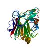

 Mass: 24.305 Da / Num. of mol.: 1 / Source method: obtained synthetically / Formula: Mg
 Mass: 18.015 Da / Num. of mol.: 150 / Source method: isolated from a natural source / Formula: H2O
 Sample preparation
 Processing