 Movie
Movie Controller

[English] 日本語
 Yorodumi
- PDB-3dgl: 1.8 A Crystal Structure of a Non-biological Protein with Bound AT... -
+ Open data

- Basic information
| Entry | Database: PDB / ID: 3dgl | ||||||
|---|---|---|---|---|---|---|---|
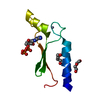
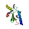
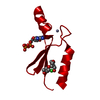
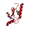
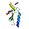
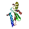
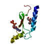
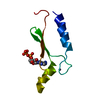
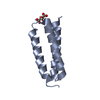
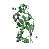
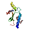
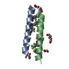
| Title | 1.8 A Crystal Structure of a Non-biological Protein with Bound ATP in a Novel Bent Conformation | ||||||
 Components Components | ATP Binding Protein-DX | ||||||
 Keywords Keywords | DE NOVO PROTEIN / alpha/beta fold / bent ATP / non-biological protein | ||||||
| Function / homology | Nuclear Transport Factor 2; Chain: A, - #210 / Nuclear Transport Factor 2; Chain: A, / Roll / Alpha Beta / ADENOSINE-5'-TRIPHOSPHATE / DI(HYDROXYETHYL)ETHER Function and homology information Function and homology information | ||||||
| Biological species | unidentified (others) | ||||||
| Method |  X-RAY DIFFRACTION / MOLECULAR REPLACEMENT / molecular replacement / Resolution: 1.8 Å X-RAY DIFFRACTION / MOLECULAR REPLACEMENT / molecular replacement / Resolution: 1.8 Å | ||||||
 Authors Authors | Simmons, C.R. / Allen, J.P. / Chaput, J.C. | ||||||
 Citation Citation | Journal: Acs Chem.Biol. / Year: 2009 Title: A synthetic protein selected for ligand binding affinity mediates ATP hydrolysis. Authors: Simmons, C.R. / Stomel, J.M. / McConnell, M.D. / Smith, D.A. / Watkins, J.L. / Allen, J.P. / Chaput, J.C. | ||||||
| History |
|
- Structure visualization
| Structure viewer | Molecule: MolmilJmol/JSmol |
|---|
- Downloads & links
-Download
| PDBx/mmCIF format | 3dgl.cif.gz | 34 KB | Display | PDBx/mmCIF format |
|---|---|---|---|---|
| PDB format | pdb3dgl.ent.gz | 21.7 KB | Display | PDB format |
| PDBx/mmJSON format | 3dgl.json.gz | Tree view | PDBx/mmJSON format | |
| Others |  Other downloads Other downloads |
-Validation report
| Arichive directory | https://data.pdbj.org/pub/pdb/validation_reports/dg/3dglftp://data.pdbj.org/pub/pdb/validation_reports/dg/3dgl | HTTPS FTP |
|---|
-Related structure data


-Links
 PDBj
PDBj
- Assembly
| Deposited unit | 
| ||||||||
|---|---|---|---|---|---|---|---|---|---|
| 1 |
| ||||||||
| Unit cell |
|
-Components
| #1: Protein | Mass: 9708.112 Da / Num. of mol.: 1 Source method: isolated from a genetically manipulated source Source: (gene. exp.) unidentified (others) / Plasmid: pIADL14 / Production host:  | ||
|---|---|---|---|
| #2: Chemical | ChemComp-ZN /   Mass: 65.409 Da / Num. of mol.: 1 / Source method: obtained synthetically / Formula: Zn Mass: 65.409 Da / Num. of mol.: 1 / Source method: obtained synthetically / Formula: Zn | ||
| #3: Chemical | ChemComp-ATP /   Mass: 507.181 Da / Num. of mol.: 1 / Source method: obtained synthetically / Formula: C10H16N5O13P3 / Comment: ATP, energy-carrying molecule*YM Mass: 507.181 Da / Num. of mol.: 1 / Source method: obtained synthetically / Formula: C10H16N5O13P3 / Comment: ATP, energy-carrying molecule*YM | ||
| #4: Chemical |   Mass: 106.120 Da / Num. of mol.: 3 / Source method: obtained synthetically / Formula: C4H10O3 Mass: 106.120 Da / Num. of mol.: 3 / Source method: obtained synthetically / Formula: C4H10O3#5: Water | ChemComp-HOH / |  Mass: 18.015 Da / Num. of mol.: 149 / Source method: isolated from a natural source / Formula: H2O Mass: 18.015 Da / Num. of mol.: 149 / Source method: isolated from a natural source / Formula: H2O |
-Experimental details
-Experiment
| Experiment | Method: X-RAY DIFFRACTION / Number of used crystals: 1 |
|---|
- Sample preparation
| Crystal | Density Matthews: 4.54 Å3/Da / Density % sol: 72.89 % |
|---|---|
| Crystal grow | Temperature: 298 K / Method: sitting drop vapor diffusion / pH: 8.5 Details: 0.1 M sodium phosphate, 0.25 M sodium citrate, 0.3 M sodium chloride, 23% polyethylene glycol 400, 0.2 M ammonium acetate, pH 8.5, sitting drop vapor diffusion, temperature 298K |
-Data collection
| Diffraction | Mean temperature: 143 K | ||||||||||||||||||||||||||||||||||||||||||||||||||||||||||||||||||
|---|---|---|---|---|---|---|---|---|---|---|---|---|---|---|---|---|---|---|---|---|---|---|---|---|---|---|---|---|---|---|---|---|---|---|---|---|---|---|---|---|---|---|---|---|---|---|---|---|---|---|---|---|---|---|---|---|---|---|---|---|---|---|---|---|---|---|---|
| Diffraction source | Source: ROTATING ANODE / Wavelength: 1.5418 Å | ||||||||||||||||||||||||||||||||||||||||||||||||||||||||||||||||||
| Detector | Type: RIGAKU RAXIS IV++ / Detector: IMAGE PLATE / Date: Oct 15, 2007 / Details: mirrors | ||||||||||||||||||||||||||||||||||||||||||||||||||||||||||||||||||
| Radiation | Protocol: SINGLE WAVELENGTH / Monochromatic (M) / Laue (L): M / Scattering type: x-ray | ||||||||||||||||||||||||||||||||||||||||||||||||||||||||||||||||||
| Radiation wavelength | Wavelength: 1.5418 Å / Relative weight: 1 | ||||||||||||||||||||||||||||||||||||||||||||||||||||||||||||||||||
| Reflection | Resolution: 1.8→25 Å / Num. obs: 16220 / % possible obs: 100 % / Redundancy: 12.9 % / Rmerge(I) obs: 0.069 / Χ2: 2.334 / Net I/σ(I): 15.5 | ||||||||||||||||||||||||||||||||||||||||||||||||||||||||||||||||||
| Reflection shell |
|
-Phasing
| Phasing | Method: molecular replacement |
|---|
- Processing
| Software |
| |||||||||||||||||||||||||||||||||||||||||||||||||||||||||||||||||
|---|---|---|---|---|---|---|---|---|---|---|---|---|---|---|---|---|---|---|---|---|---|---|---|---|---|---|---|---|---|---|---|---|---|---|---|---|---|---|---|---|---|---|---|---|---|---|---|---|---|---|---|---|---|---|---|---|---|---|---|---|---|---|---|---|---|---|
| Refinement | Method to determine structure: MOLECULAR REPLACEMENT / Resolution: 1.8→25 Å / Cor.coef. Fo:Fc: 0.958 / Cor.coef. Fo:Fc free: 0.94 / Occupancy max: 1 / Occupancy min: 0.5 / SU B: 1.57 / SU ML: 0.05 / Cross valid method: THROUGHOUT / σ(F): 0 / ESU R: 0.08 / ESU R Free: 0.083 / Stereochemistry target values: MAXIMUM LIKELIHOOD / Details: HYDROGENS HAVE BEEN ADDED IN THE RIDING POSITIONS
| |||||||||||||||||||||||||||||||||||||||||||||||||||||||||||||||||
| Solvent computation | Ion probe radii: 0.8 Å / Shrinkage radii: 0.8 Å / VDW probe radii: 1.2 Å / Solvent model: MASK | |||||||||||||||||||||||||||||||||||||||||||||||||||||||||||||||||
| Displacement parameters | Biso max: 101.24 Å2 / Biso mean: 24.359 Å2 / Biso min: 11.01 Å2
| |||||||||||||||||||||||||||||||||||||||||||||||||||||||||||||||||
| Refinement step | Cycle: LAST / Resolution: 1.8→25 Å
| |||||||||||||||||||||||||||||||||||||||||||||||||||||||||||||||||
| Refine LS restraints |
| |||||||||||||||||||||||||||||||||||||||||||||||||||||||||||||||||
| LS refinement shell | Resolution: 1.8→1.849 Å / Total num. of bins used: 20
|
