 Movie
Movie Controller

[English] 日本語
 Yorodumi
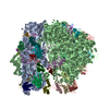
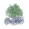
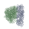
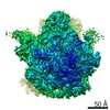
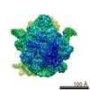
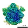
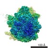
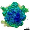
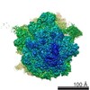
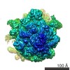
- PDB-3dg0: Coordinates of 16S and 23S rRNAs fitted into the cryo-EM map of E... -
+ Open data

- Basic information
| Entry | Database: PDB / ID: 3dg0 | ||||||
|---|---|---|---|---|---|---|---|
| Title | Coordinates of 16S and 23S rRNAs fitted into the cryo-EM map of EF-G-bound translocation complex | ||||||
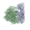
 Components Components |
| ||||||
 Keywords Keywords | RIBOSOME / Ribosome EF-G ratchet motion | ||||||
| Function / homology | RNA / RNA (> 10) / RNA (> 100) / RNA (> 1000) Function and homology information Function and homology information | ||||||
| Biological species |  | ||||||
| Method | ELECTRON MICROSCOPY / single particle reconstruction / cryo EM / Resolution: 10.8 Å | ||||||
 Authors Authors | Gao, H. / LeBarron, J. / Frank, J. | ||||||
 Citation Citation | Journal: To be published Title: Ribosomal Dynamics: Intrinsic Instability of a Moleculaar Machine Authors: Gao, H. / LeBarron, J. / Frank, J. | ||||||
| History |
|
- Structure visualization
| Movie |
Movie viewer |
|---|---|
| Structure viewer | Molecule: MolmilJmol/JSmol |
- Downloads & links
-Download
| PDBx/mmCIF format | 3dg0.cif.gz | 140.4 KB | Display | PDBx/mmCIF format |
|---|---|---|---|---|
| PDB format | pdb3dg0.ent.gz | 82 KB | Display | PDB format |
| PDBx/mmJSON format | 3dg0.json.gz | Tree view | PDBx/mmJSON format | |
| Others |  Other downloads Other downloads |
-Validation report
| Arichive directory | https://data.pdbj.org/pub/pdb/validation_reports/dg/3dg0ftp://data.pdbj.org/pub/pdb/validation_reports/dg/3dg0 | HTTPS FTP |
|---|
-Related structure data
| Related structure data | 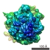 1363M 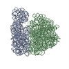 3dg2C 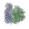 3dg4C 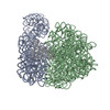 3dg5C C: citing same article ( M: map data used to model this data |
|---|---|
| Similar structure data |
-Links
 PDBj
PDBj


- Assembly
| Deposited unit | 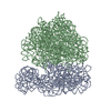
|
|---|---|
| 1 |
|
-Components
| #1: RNA chain | Mass: 499690.031 Da / Num. of mol.: 1 / Source method: isolated from a natural source / Source: (natural) |
|---|---|
| #2: RNA chain | Mass: 941612.375 Da / Num. of mol.: 1 / Source method: isolated from a natural source / Source: (natural) |
| Sequence details | THE STRUCTURE CONTAINS P ATOMS ONLY |
-Experimental details
-Experiment
| Experiment | Method: ELECTRON MICROSCOPY |
|---|---|
| EM experiment | Aggregation state: PARTICLE / 3D reconstruction method: single particle reconstruction |
- Sample preparation
| Component | Name: E. coli 70S ribosome -EF-G-GNPNP / Type: RIBOSOME |
|---|---|
| Buffer solution | Name: polymix / pH: 7.5 / Details: polymix |
| Specimen | Conc.: 32 mg/ml / Embedding applied: NO / Shadowing applied: NO / Staining applied: NO / Vitrification applied: YES |
| Vitrification | Cryogen name: ETHANE / Details: Rapid freezing in liquid ethane |
- Electron microscopy imaging
| Experimental equipment |  Model: Tecnai F20 / Image courtesy: FEI Company |
|---|---|
| Microscopy | Model: FEI TECNAI F20 / Date: Jul 11, 2003 |
| Electron gun | Electron source:  FIELD EMISSION GUN / Accelerating voltage: 200 kV / Illumination mode: FLOOD BEAM FIELD EMISSION GUN / Accelerating voltage: 200 kV / Illumination mode: FLOOD BEAM |
| Electron lens | Mode: BRIGHT FIELD / Nominal magnification: 50000 X / Calibrated magnification: 49696 X / Nominal defocus max: 4000 nm / Nominal defocus min: 2000 nm / Cs: 2 mm |
| Specimen holder | Temperature: 93 K / Tilt angle max: 0 ° / Tilt angle min: 0 ° |
| Image recording | Electron dose: 20 e/Å2 / Film or detector model: KODAK SO-163 FILM |
- Processing
| EM software |
| |||||||||||||||||||||
|---|---|---|---|---|---|---|---|---|---|---|---|---|---|---|---|---|---|---|---|---|---|---|
| CTF correction | Details: CTF correction of 3D map | |||||||||||||||||||||
| Symmetry | Point symmetry: C1 (asymmetric) | |||||||||||||||||||||
| 3D reconstruction | Method: reference-based alignment / Resolution: 10.8 Å / Nominal pixel size: 2.8 Å / Actual pixel size: 2.76 Å / Magnification calibration: TMV / Details: SPIDER package / Symmetry type: POINT | |||||||||||||||||||||
| Atomic model building | Protocol: RIGID BODY FIT / Space: REAL Target criteria: cross-correlation coefficient, real-space R factor Details: METHOD--auto REFINEMENT PROTOCOL--multi-rigid body, real-space refinement | |||||||||||||||||||||
| Atomic model building |
| |||||||||||||||||||||
| Refinement step | Cycle: LAST
|

