 Movie
Movie Controller


+ Open data

- Basic information
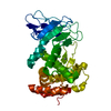
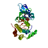
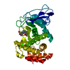
| Entry | Database: PDB / ID: 3dbk | ||||||
|---|---|---|---|---|---|---|---|
| Title | Pseudomonas aeruginosa elastase with phosphoramidon | ||||||
 Components Components | Elastase | ||||||
 Keywords Keywords | HYDROLASE / zinc metalloprotease / enzyme / phosphoramidon / protease inhibitor complex / Calcium / Metal-binding / Metalloprotease / Protease / Secreted / Zinc / Zymogen | ||||||
| Function / homology |  Function and homology information Function and homology informationpseudolysin / protein transport by the Sec complex / protein secretion by the type II secretion system / single-species biofilm formation / bacterial-type flagellum-dependent swarming motility / metalloendopeptidase activity / endopeptidase activity / proteolysis / extracellular region / metal ion binding Similarity search - Function | ||||||
| Biological species |   Pseudomonas aeruginosa (bacteria) Pseudomonas aeruginosa (bacteria) | ||||||
| Method |  X-RAY DIFFRACTION / SYNCHROTRON / Resolution: 1.4 Å X-RAY DIFFRACTION / SYNCHROTRON / Resolution: 1.4 Å | ||||||
 Authors Authors | McKay, D.B. / Overgaard, M.T. | ||||||
 Citation Citation | Journal: To be Published Title: Structure of the Elastase of Pseudomonas aeruginosa Complexed with Phosphoramidon Authors: Overgaard, M.T. / McKay, D.B. #1: Journal: J.Biol.Chem. / Year: 1991Title: Three-Dimensional Structure of the Elastase of Pseudomonas aeruginosa at 1.5 Angstrom Resolution Authors: Thayer, M.M. / Flaherty, K.M. / McKay, D.B. #2: Journal: Biochemistry / Year: 1992Title: Structural comparison suggests that thermolysin and related neutral proteases undergo hinge-bending motion during catalysis Authors: Holland, D.R. / Tronrud, D.E. / Pley, H.W. / Flaherty, K.M. / Stark, W. / Jansonius, J.N. / McKay, D.B. / Matthews, B.W. | ||||||
| History |
|
- Structure visualization
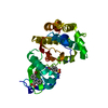
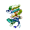
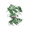
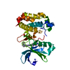
| Structure viewer | Molecule: MolmilJmol/JSmol |
|---|
- Downloads & links
-Download
| PDBx/mmCIF format | 3dbk.cif.gz | 82.8 KB | Display | PDBx/mmCIF format |
|---|---|---|---|---|
| PDB format | pdb3dbk.ent.gz | 60.1 KB | Display | PDB format |
| PDBx/mmJSON format | 3dbk.json.gz | Tree view | PDBx/mmJSON format | |
| Others |  Other downloads Other downloads |
-Validation report
| Arichive directory | https://data.pdbj.org/pub/pdb/validation_reports/db/3dbkftp://data.pdbj.org/pub/pdb/validation_reports/db/3dbk | HTTPS FTP |
|---|
-Related structure data
| Related structure data |  1u4gS S: Starting model for refinement |
|---|---|
| Similar structure data |


-Links
 PDBj
PDBj- Assembly
| Deposited unit | 
| ||||||||
|---|---|---|---|---|---|---|---|---|---|
| 1 |
| ||||||||
| Unit cell |
|
-Components
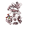
-Protein , 1 types, 1 molecules A
| #1: Protein | Mass: 33175.531 Da / Num. of mol.: 1 / Source method: isolated from a natural source Details: Isolated as mature extracellular protease from Pseudomonas aeruginosa cultures Source: (natural) Pseudomonas aeruginosa (bacteria) / References: UniProt: P14756 |
|---|
-Non-polymers , 5 types, 382 molecules 


| #2: Chemical | ChemComp-ZN /  Mass: 65.409 Da / Num. of mol.: 1 / Source method: obtained synthetically / Formula: Zn Mass: 65.409 Da / Num. of mol.: 1 / Source method: obtained synthetically / Formula: Zn | ||||
|---|---|---|---|---|---|
| #3: Chemical | ChemComp-CA /  Mass: 40.078 Da / Num. of mol.: 1 / Source method: obtained synthetically / Formula: Ca Mass: 40.078 Da / Num. of mol.: 1 / Source method: obtained synthetically / Formula: Ca | ||||
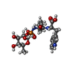
| #4: Chemical |  Mass: 96.063 Da / Num. of mol.: 3 / Source method: obtained synthetically / Formula: SO4 Mass: 96.063 Da / Num. of mol.: 3 / Source method: obtained synthetically / Formula: SO4#5: Chemical | ChemComp-RDF / |  Type: peptide-like, Glycopeptide / Class: Enzyme inhibitor / Mass: 543.504 Da / Num. of mol.: 1 / Source method: obtained synthetically / Formula: C23H34N3O10P / References: PHOSPHORAMIDON Type: peptide-like, Glycopeptide / Class: Enzyme inhibitor / Mass: 543.504 Da / Num. of mol.: 1 / Source method: obtained synthetically / Formula: C23H34N3O10P / References: PHOSPHORAMIDON#6: Water | ChemComp-HOH / | Mass: 18.015 Da / Num. of mol.: 376 / Source method: isolated from a natural source / Formula: H2O |
-Details
| Has protein modification | Y |
|---|
-Experimental details
-Experiment
| Experiment | Method: X-RAY DIFFRACTION / Number of used crystals: 1 |
|---|
- Sample preparation
| Crystal | Density Matthews: 2.05 Å3/Da / Density % sol: 40.1 % |
|---|---|
| Crystal grow | Temperature: 298 K / Method: vapor diffusion, hanging drop / pH: 6.5 Details: crystallized from 1.2-1.7 M ammonium sulfate, 0.1 M MOPS buffer, 1 micromolar Ca2+, 1 micromolar phosphoramidon, pH 6.5, VAPOR DIFFUSION, HANGING DROP, temperature 298K |
-Data collection
| Diffraction | Mean temperature: 100 K |
|---|---|
| Diffraction source | Source: SYNCHROTRON / Site: SSRL  / Beamline: BL11-1 / Wavelength: 0.98 Å / Beamline: BL11-1 / Wavelength: 0.98 Å |
| Detector | Type: ADSC QUANTUM 315 / Detector: CCD / Date: Dec 6, 2001 |
| Radiation | Protocol: SINGLE WAVELENGTH / Monochromatic (M) / Laue (L): M / Scattering type: x-ray |
| Radiation wavelength | Wavelength: 0.98 Å / Relative weight: 1 |
| Reflection | Resolution: 1.4→50 Å / Num. all: 54472 / Num. obs: 50494 / % possible obs: 92.7 % / Observed criterion σ(F): 0 / Observed criterion σ(I): 0 / Redundancy: 3.1 % / Biso Wilson estimate: 12 Å2 / Rsym value: 0.065 / Net I/σ(I): 7.6 |
| Reflection shell | Resolution: 1.4→1.43 Å / Redundancy: 2 % / Mean I/σ(I) obs: 4.7 / Rsym value: 0.134 / % possible all: 92.9 |
- Processing
| Software |
| ||||||||||||||||||||||||||||||||||||||||||||||||||||||||||||||||||||||||||||||||
|---|---|---|---|---|---|---|---|---|---|---|---|---|---|---|---|---|---|---|---|---|---|---|---|---|---|---|---|---|---|---|---|---|---|---|---|---|---|---|---|---|---|---|---|---|---|---|---|---|---|---|---|---|---|---|---|---|---|---|---|---|---|---|---|---|---|---|---|---|---|---|---|---|---|---|---|---|---|---|---|---|---|
| Refinement | Starting model: 1U4G Resolution: 1.4→29.17 Å / Rfactor Rfree error: 0.004 / Data cutoff high absF: 1793957.18 / Data cutoff low absF: 0 / Isotropic thermal model: RESTRAINED / Cross valid method: THROUGHOUT / σ(F): 0 / Stereochemistry target values: Engh & Huber / Details: BULK SOLVENT MODEL USED
| ||||||||||||||||||||||||||||||||||||||||||||||||||||||||||||||||||||||||||||||||
| Solvent computation | Solvent model: FLAT MODEL / Bsol: 41.8855 Å2 / ksol: 0.4 e/Å3 | ||||||||||||||||||||||||||||||||||||||||||||||||||||||||||||||||||||||||||||||||
| Displacement parameters | Biso mean: 14.5 Å2
| ||||||||||||||||||||||||||||||||||||||||||||||||||||||||||||||||||||||||||||||||
| Refine analyze |
| ||||||||||||||||||||||||||||||||||||||||||||||||||||||||||||||||||||||||||||||||
| Refinement step | Cycle: LAST / Resolution: 1.4→29.17 Å
| ||||||||||||||||||||||||||||||||||||||||||||||||||||||||||||||||||||||||||||||||
| Refine LS restraints |
| ||||||||||||||||||||||||||||||||||||||||||||||||||||||||||||||||||||||||||||||||
| LS refinement shell | Resolution: 1.4→1.45 Å / Rfactor Rfree error: 0.016 / Total num. of bins used: 10
| ||||||||||||||||||||||||||||||||||||||||||||||||||||||||||||||||||||||||||||||||
| Xplor file |
|
