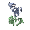
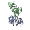
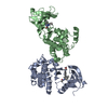
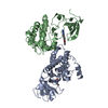
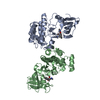
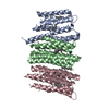
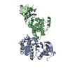
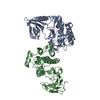
[English] 日本語
Yorodumi
- PDB-3d7u: Structural basis for the recognition of c-Src by its inactivator Csk -

+
Open data


ID or keywords:

Loading...

-
Basic information

Entry
Database: PDB / ID: 3d7u
TitleStructural basis for the recognition of c-Src by its inactivator Csk
Components
  • Proto-oncogene tyrosine-protein kinase Src
  • Tyrosine-protein kinase CSK
KeywordsTRANSFERASE / Csk c-Src Tyrosine kinase / ATP-binding / Kinase / Membrane / Nucleotide-binding / Phosphoprotein / SH2 domain / SH3 domain / Tyrosine-protein kinase / Lipoprotein / Myristate / Proto-oncogene
Function / homology
Function and homology information


negative regulation of Golgi to plasma membrane protein transport / regulation of Fc receptor mediated stimulatory signaling pathway / negative regulation of low-density lipoprotein particle clearance / Signaling by ERBB2 / Nuclear signaling by ERBB4 / Signaling by SCF-KIT / Regulation of KIT signaling / Signaling by EGFR / GAB1 signalosome / Regulation of gap junction activity ...negative regulation of Golgi to plasma membrane protein transport / regulation of Fc receptor mediated stimulatory signaling pathway / negative regulation of low-density lipoprotein particle clearance / Signaling by ERBB2 / Nuclear signaling by ERBB4 / Signaling by SCF-KIT / Regulation of KIT signaling / Signaling by EGFR / GAB1 signalosome / Regulation of gap junction activity / FCGR activation / PECAM1 interactions / Co-stimulation by CD28 / Co-inhibition by CTLA4 / EPHA-mediated growth cone collapse / Ephrin signaling / G alpha (i) signalling events / GP1b-IX-V activation signalling / Thrombin signalling through proteinase activated receptors (PARs) / VEGFR2 mediated cell proliferation / RET signaling / Receptor Mediated Mitophagy / ADP signalling through P2Y purinoceptor 1 / RAF activation / PIP3 activates AKT signaling / EPH-ephrin mediated repulsion of cells / PI5P, PP2A and IER3 Regulate PI3K/AKT Signaling / Activated NTRK3 signals through PI3K / Downstream signal transduction / Downregulation of ERBB4 signaling / Cyclin D associated events in G1 / Regulation of RUNX3 expression and activity / MAP2K and MAPK activation / Integrin signaling / GRB2:SOS provides linkage to MAPK signaling for Integrins / DCC mediated attractive signaling / MET activates PTK2 signaling / Extra-nuclear estrogen signaling / EPHB-mediated forward signaling / p130Cas linkage to MAPK signaling for integrins / VEGFA-VEGFR2 Pathway / connexin binding / adherens junction organization / proline-rich region binding / negative regulation of bone resorption / cellular response to peptide hormone stimulus / negative regulation of phagocytosis / Phosphorylation of CD3 and TCR zeta chains / protein kinase A catalytic subunit binding / negative regulation of intrinsic apoptotic signaling pathway / oligodendrocyte differentiation / negative regulation of interleukin-6 production / RHOH GTPase cycle / immune system process / Co-inhibition by PD-1 / GAB1 signalosome / progesterone receptor signaling pathway / T cell costimulation / Integrin signaling / Negative regulation of FLT3 / protein tyrosine kinase binding / negative regulation of extrinsic apoptotic signaling pathway / non-membrane spanning protein tyrosine kinase activity / non-specific protein-tyrosine kinase / Signaling by high-kinase activity BRAF mutants / MAP2K and MAPK activation / negative regulation of ERK1 and ERK2 cascade / epidermal growth factor receptor signaling pathway / Signaling by RAF1 mutants / Signaling by moderate kinase activity BRAF mutants / Paradoxical activation of RAF signaling by kinase inactive BRAF / Signaling downstream of RAS mutants / Signaling by BRAF and RAF1 fusions / cell junction / cell-cell junction / T cell receptor signaling pathway / protein tyrosine kinase activity / protein phosphatase binding / adaptive immune response / cell differentiation / cytoskeleton / mitochondrial inner membrane / regulation of cell cycle / cell adhesion / endosome membrane / protein phosphorylation / signaling receptor binding / negative regulation of cell population proliferation / focal adhesion / heme binding / perinuclear region of cytoplasm / protein-containing complex / extracellular exosome / ATP binding / metal ion binding / identical protein binding / nucleus / membrane / plasma membrane / cytosol
Similarity search - Function
CSK-like, SH2 domain / : / SH3 domain / SH2 domain / Src homology 2 (SH2) domain profile. / Src homology 2 domains / SH2 domain / SH2 domain superfamily / Src homology 3 domains / SH3-like domain superfamily ...CSK-like, SH2 domain / : / SH3 domain / SH2 domain / Src homology 2 (SH2) domain profile. / Src homology 2 domains / SH2 domain / SH2 domain superfamily / Src homology 3 domains / SH3-like domain superfamily / Src homology 3 (SH3) domain profile. / SH3 domain / Tyrosine-protein kinase, catalytic domain / Tyrosine kinase, catalytic domain / Tyrosine protein kinases specific active-site signature. / Tyrosine-protein kinase, active site / Serine-threonine/tyrosine-protein kinase, catalytic domain / Protein tyrosine and serine/threonine kinase / Protein kinase, ATP binding site / Protein kinases ATP-binding region signature. / Protein kinase domain profile. / Protein kinase domain / Protein kinase-like domain superfamily
Similarity search - Domain/homology
Proto-oncogene tyrosine-protein kinase Src / Tyrosine-protein kinase CSK
Similarity search - Component
Biological speciesHomo sapiens (human)
Gallus gallus (chicken)
MethodX-RAY DIFFRACTION / SYNCHROTRON / MOLECULAR REPLACEMENT / Resolution: 4.111 Å
AuthorsLevinson, N.M. / Seeliger, M.A. / Cole, P.A. / Kuriyan, J.
CitationJournal: Cell(Cambridge,Mass.) / Year: 2008
Title: Structural basis for the recognition of c-Src by its inactivator Csk.
Authors: Levinson, N.M. / Seeliger, M.A. / Cole, P.A. / Kuriyan, J.
History
DepositionMay 21, 2008Deposition site: RCSB / Processing site: RCSB
Revision 1.0Aug 5, 2008Provider: repository / Type: Initial release
Revision 1.1Jul 13, 2011Group: Version format compliance
Revision 1.2Sep 18, 2013Group: Derived calculations
Revision 1.3Feb 21, 2024Group: Data collection / Database references
Category: chem_comp_atom / chem_comp_bond ...chem_comp_atom / chem_comp_bond / database_2 / struct_ref_seq_dif
Item: _database_2.pdbx_DOI / _database_2.pdbx_database_accession / _struct_ref_seq_dif.details

-
Structure visualization

Structure viewerMolecule:
MolmilJmol/JSmol

Downloads & links

-
Assembly

Deposited unit
A: Tyrosine-protein kinase CSK
B: Proto-oncogene tyrosine-protein kinase Src
C: Tyrosine-protein kinase CSK
D: Proto-oncogene tyrosine-protein kinase Src


Theoretical massNumber of molelcules
Total (without water)122,8524
Polymers122,8524
Non-polymers00
Water00
1
A: Tyrosine-protein kinase CSK
B: Proto-oncogene tyrosine-protein kinase Src


Theoretical massNumber of molelcules
Total (without water)61,4262
Polymers61,4262
Non-polymers00
Water0
TypeNameSymmetry operationNumber
identity operation1_555x,y,z1
2
C: Tyrosine-protein kinase CSK
D: Proto-oncogene tyrosine-protein kinase Src


Theoretical massNumber of molelcules
Total (without water)61,4262
Polymers61,4262
Non-polymers00
Water0
TypeNameSymmetry operationNumber
identity operation1_555x,y,z1
Unit cell
Length a, b, c (Å)135.839, 135.839, 129.509
Angle α, β, γ (deg.)90.00, 90.00, 120.00
Int Tables number144
Space group name H-MP31

-
Components

#1: Protein Tyrosine-protein kinase CSK / C-SRC kinase / Protein-tyrosine kinase CYL


Mass: 29624.256 Da / Num. of mol.: 2 / Fragment: Csk kinase domain
Source method: isolated from a genetically manipulated source
Source: (gene. exp.) Homo sapiens (human) / Gene: CSK / Production host: Escherichia coli (E. coli)
References: UniProt: P41240, non-specific protein-tyrosine kinase
#2: Protein Proto-oncogene tyrosine-protein kinase Src / pp60c-src / p60-Src / c-Src


Mass: 31801.605 Da / Num. of mol.: 2 / Fragment: c-Src kinase domain
Source method: isolated from a genetically manipulated source
Source: (gene. exp.) Gallus gallus (chicken) / Gene: SRC / Production host: Escherichia coli (E. coli)
References: UniProt: P00523, non-specific protein-tyrosine kinase

-
Experimental details

-
Experiment

ExperimentMethod: X-RAY DIFFRACTION

-
Sample preparation

CrystalDensity Matthews: 5.62 Å3/Da / Density % sol: 78.1 %

-
Data collection

Diffraction sourceSource: SYNCHROTRON / Site: ALS / Beamline: 8.2.1
RadiationProtocol: SINGLE WAVELENGTH / Monochromatic (M) / Laue (L): M / Scattering type: x-ray
Radiation wavelengthRelative weight: 1
ReflectionResolution: 4.0117→46.8705 Å / Num. obs: 22041

-
Processing

Software
NameVersionClassification
PHENIX(phenix.refine)refinement
HKL-2000data reduction
HKL-2000data scaling
RefinementMethod to determine structure: MOLECULAR REPLACEMENT / Resolution: 4.111→46.866 Å / Occupancy max: 1 / Occupancy min: 1 / SU ML: 0.67 / σ(F): 1.96 / Phase error: 31.85 / Stereochemistry target values: ML
RfactorNum. reflection% reflection
Rfree0.302 1053 5.14 %
Rwork0.294 19446 -
obs0.294 20499 98.54 %
Solvent computationShrinkage radii: 0.9 Å / VDW probe radii: 1.11 Å / Solvent model: FLAT BULK SOLVENT MODEL / Bsol: 123.071 Å2 / ksol: 0.334 e/Å3
Displacement parametersBiso max: 271.05 Å2 / Biso mean: 191.051 Å2 / Biso min: 119.61 Å2
Baniso -1Baniso -2Baniso -3
1-16.564 Å2-0 Å2-0 Å2
2--16.564 Å2-0 Å2
3----33.129 Å2
Refinement stepCycle: LAST / Resolution: 4.111→46.866 Å
ProteinNucleic acidLigandSolventTotal
Num. atoms8153 0 0 0 8153
Refine LS restraints
Refine-IDTypeDev idealNumber
X-RAY DIFFRACTIONf_bond_d0.028341
X-RAY DIFFRACTIONf_angle_d1.59211281
X-RAY DIFFRACTIONf_dihedral_angle_d21.6953102
X-RAY DIFFRACTIONf_chiral_restr0.0881232
X-RAY DIFFRACTIONf_plane_restr0.0081440
LS refinement shell

Refine-ID: X-RAY DIFFRACTION / Total num. of bins used: 8

Resolution (Å)Rfactor RfreeNum. reflection RfreeRfactor RworkNum. reflection RworkNum. reflection all% reflection obs (%)
4.111-4.2980.3661510.3632431258298
4.298-4.5240.2981280.3432436256499
4.524-4.8070.2861170.31924872604100
4.807-5.1780.3441330.3092409254299
5.178-5.6990.3511420.332463260599
5.699-6.5210.3141410.3072416255799
6.521-8.2090.2721280.2732408253698
8.209-46.8690.2471130.2362396250996

+
About Yorodumi

-
News

-
Feb 9, 2022. New format data for meta-information of EMDB entries

New format data for meta-information of EMDB entries

  • Version 3 of the EMDB header file is now the official format.
  • The previous official version 1.9 will be removed from the archive.

Related info.:EMDB header

External links:wwPDB to switch to version 3 of the EMDB data model

-
Aug 12, 2020. Covid-19 info

Covid-19 info

URL: https://pdbj.org/emnavi/covid19.php

New page: Covid-19 featured information page in EM Navigator.

Related info.:Covid-19 info / Mar 5, 2020. Novel coronavirus structure data

+
Mar 5, 2020. Novel coronavirus structure data

Novel coronavirus structure data

Related info.:Yorodumi Speices / Aug 12, 2020. Covid-19 info

External links:COVID-19 featured content - PDBj / Molecule of the Month (242):Coronavirus Proteases

+
Jan 31, 2019. EMDB accession codes are about to change! (news from PDBe EMDB page)

EMDB accession codes are about to change! (news from PDBe EMDB page)

  • The allocation of 4 digits for EMDB accession codes will soon come to an end. Whilst these codes will remain in use, new EMDB accession codes will include an additional digit and will expand incrementally as the available range of codes is exhausted. The current 4-digit format prefixed with “EMD-” (i.e. EMD-XXXX) will advance to a 5-digit format (i.e. EMD-XXXXX), and so on. It is currently estimated that the 4-digit codes will be depleted around Spring 2019, at which point the 5-digit format will come into force.
  • The EM Navigator/Yorodumi systems omit the EMD- prefix.

Related info.:Q: What is EMD? / ID/Accession-code notation in Yorodumi/EM Navigator

External links:EMDB Accession Codes are Changing Soon! / Contact to PDBj

+
Jul 12, 2017. Major update of PDB

Major update of PDB

  • wwPDB released updated PDB data conforming to the new PDBx/mmCIF dictionary.
  • This is a major update changing the version number from 4 to 5, and with Remediation, in which all the entries are updated.
  • In this update, many items about electron microscopy experimental information are reorganized (e.g. em_software).
  • Now, EM Navigator and Yorodumi are based on the updated data.

External links:wwPDB Remediation / Enriched Model Files Conforming to OneDep Data Standards Now Available in the PDB FTP Archive

-
Yorodumi

Thousand views of thousand structures

  • Yorodumi is a browser for structure data from EMDB, PDB, SASBDB, etc.
  • This page is also the successor to EM Navigator detail page, and also detail information page/front-end page for Omokage search.
  • The word "yorodu" (or yorozu) is an old Japanese word meaning "ten thousand". "mi" (miru) is to see.

Related info.:EMDB / PDB / SASBDB / Comparison of 3 databanks / Yorodumi Search / Aug 31, 2016. New EM Navigator & Yorodumi / Yorodumi Papers / Jmol/JSmol / Function and homology information / Changes in new EM Navigator and Yorodumi

Read more