 Movie
Movie Controller

[English] 日本語
 Yorodumi
- PDB-3d72: 1.65 Angstrom crystal structure of the Cys71Val variant in the fu... -
+ Open data

- Basic information
| Entry | Database: PDB / ID: 3d72 | ||||||
|---|---|---|---|---|---|---|---|
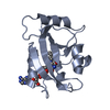
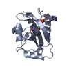
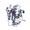
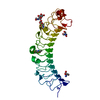
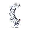
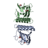
| Title | 1.65 Angstrom crystal structure of the Cys71Val variant in the fungal photoreceptor VVD | ||||||
 Components Components | Vivid PAS protein VVD | ||||||
 Keywords Keywords | SIGNALING PROTEIN / Circadian / photoreceptor / blue-light / LOV / PAS / VVD | ||||||
| Function / homology |  Function and homology information Function and homology information | ||||||
| Biological species |  Neurospora crassa (fungus) Neurospora crassa (fungus) | ||||||
| Method |  X-RAY DIFFRACTION / SYNCHROTRON / MOLECULAR REPLACEMENT / Resolution: 1.65 Å X-RAY DIFFRACTION / SYNCHROTRON / MOLECULAR REPLACEMENT / Resolution: 1.65 Å | ||||||
 Authors Authors | Zoltowski, B.D. / Crane, B.R. | ||||||
 Citation Citation | Journal: Biochemistry / Year: 2008 Title: Light activation of the LOV protein vivid generates a rapidly exchanging dimer. Authors: Zoltowski, B.D. / Crane, B.R. | ||||||
| History |
|
- Structure visualization
| Structure viewer | Molecule: MolmilJmol/JSmol |
|---|
- Downloads & links
-Download
| PDBx/mmCIF format | 3d72.cif.gz | 86 KB | Display | PDBx/mmCIF format |
|---|---|---|---|---|
| PDB format | pdb3d72.ent.gz | 63.8 KB | Display | PDB format |
| PDBx/mmJSON format | 3d72.json.gz | Tree view | PDBx/mmJSON format | |
| Others |  Other downloads Other downloads |
-Validation report
| Summary document | 3d72_validation.pdf.gz | 996.2 KB | Display | wwPDB validaton report |
|---|---|---|---|---|
| Full document | 3d72_full_validation.pdf.gz | 1005.4 KB | Display | |
| Data in XML | 3d72_validation.xml.gz | 20.6 KB | Display | |
| Data in CIF | 3d72_validation.cif.gz | 29.7 KB | Display | |
| Arichive directory | https://data.pdbj.org/pub/pdb/validation_reports/d7/3d72ftp://data.pdbj.org/pub/pdb/validation_reports/d7/3d72 | HTTPS FTP |
-Related structure data
| Related structure data | 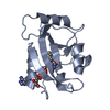 2pd7S S: Starting model for refinement |
|---|---|
| Similar structure data |


-Links
 PDBj
PDBj

- Assembly
| Deposited unit | 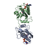
| ||||||||
|---|---|---|---|---|---|---|---|---|---|
| 1 |
| ||||||||
| Unit cell |
|
-Components
| #1: Protein | Mass: 16996.496 Da / Num. of mol.: 2 / Fragment: Fungal Photoreceptor VVD / Mutation: c71v Source method: isolated from a genetically manipulated source Source: (gene. exp.) Neurospora crassa (fungus) / Gene: vvd, G17A4.050 / Plasmid: pET28b / Production host:  #2: Chemical |   Mass: 785.550 Da / Num. of mol.: 2 / Source method: obtained synthetically / Formula: C27H33N9O15P2 / Comment: FAD*YM Mass: 785.550 Da / Num. of mol.: 2 / Source method: obtained synthetically / Formula: C27H33N9O15P2 / Comment: FAD*YM#3: Water | ChemComp-HOH / |  Mass: 18.015 Da / Num. of mol.: 462 / Source method: isolated from a natural source / Formula: H2O Mass: 18.015 Da / Num. of mol.: 462 / Source method: isolated from a natural source / Formula: H2O |
|---|
-Experimental details
-Experiment
| Experiment | Method: X-RAY DIFFRACTION / Number of used crystals: 1 |
|---|
- Sample preparation
| Crystal | Density Matthews: 2.47 Å3/Da / Density % sol: 50.14 % |
|---|---|
| Crystal grow | Temperature: 295 K / Method: vapor diffusion, hanging drop / pH: 5.6 Details: 26% PEG 4000, 100 mM triSodium Citrate pH 5.6, 100 mM Sodium Acetate, VAPOR DIFFUSION, HANGING DROP, temperature 295K |
-Data collection
| Diffraction | Mean temperature: 77 K |
|---|---|
| Diffraction source | Source: SYNCHROTRON / Site: CHESS  / Beamline: F1 / Wavelength: 0.9179 Å / Beamline: F1 / Wavelength: 0.9179 Å |
| Detector | Type: ADSC QUANTUM 270 / Detector: CCD / Date: Oct 27, 2007 |
| Radiation | Monochromator: Horizontal bent Si(111), asymmetrically cut with water cooled Cu Block Protocol: SINGLE WAVELENGTH / Monochromatic (M) / Laue (L): M / Scattering type: x-ray |
| Radiation wavelength | Wavelength: 0.9179 Å / Relative weight: 1 |
| Reflection | Resolution: 1.65→50 Å / Num. all: 39657 / Num. obs: 39115 / % possible obs: 98.6 % / Observed criterion σ(F): 0 / Observed criterion σ(I): 0 / Redundancy: 4.1 % / Biso Wilson estimate: 16.93 Å2 / Rmerge(I) obs: 0.063 / Net I/σ(I): 37.95 |
| Reflection shell | Resolution: 1.65→1.71 Å / Redundancy: 3.7 % / Rmerge(I) obs: 0.18 / Mean I/σ(I) obs: 6.9 / Num. unique all: 3682 / % possible all: 94.1 |
- Processing
| Software |
| |||||||||||||||||||||||||
|---|---|---|---|---|---|---|---|---|---|---|---|---|---|---|---|---|---|---|---|---|---|---|---|---|---|---|
| Refinement | Method to determine structure: MOLECULAR REPLACEMENT Starting model: 2PD7 Resolution: 1.65→50 Å / σ(F): 0 / Stereochemistry target values: Engh & Huber
| |||||||||||||||||||||||||
| Refinement step | Cycle: LAST / Resolution: 1.65→50 Å
| |||||||||||||||||||||||||
| Refine LS restraints |
|
