Mass: 18.015 Da / Num. of mol.: 20 / Source method: isolated from a natural source / Formula: H2O
Has protein modification
N
-
Experimental details
-
Experiment
Experiment
Method: X-RAY DIFFRACTION / Number of used crystals: 1
-
Sample preparation
Crystal
Density Matthews: 2.26 Å3/Da / Density % sol: 45.48 % Description: The structure factor file contains Friedel pairs
Crystal grow
Temperature: 292 K / Method: vapor diffusion, sitting drop Details: Protein solution: 20 mM Tris-HCl pH 7.5, 100 mM NaCl, 5 mM Acetyl-CoA. Reservoir solution: 200 mM Ammonium citrate pH 5.0, 20% PEG 3350, VAPOR DIFFUSION, SITTING DROP, temperature 292K
Resolution: 2.8→2.9 Å / Redundancy: 3.1 % / Rmerge(I) obs: 0.354 / Mean I/σ(I) obs: 2.25 / Rsym value: 0.324 / % possible all: 100
-
Processing
Software
Name
Version
Classification
REFMAC
5.2.0003
refinement
ADSC
Quantum
datacollection
DENZO
datareduction
SCALEPACK
datascaling
SnB
phasing
SOLVE
phasing
RESOLVE
phasing
Refinement
Method to determine structure: SAD / Resolution: 2.8→29.35 Å / Cor.coef. Fo:Fc: 0.923 / Cor.coef. Fo:Fc free: 0.88 / SU B: 34.071 / SU ML: 0.302 / TLS residual ADP flag: LIKELY RESIDUAL / Cross valid method: THROUGHOUT / ESU R Free: 0.406 / Stereochemistry target values: MAXIMUM LIKELIHOOD Details: The Friedel pairs were used in phasing. Program CNS 1.2 has also been used in refinement
Rfactor
Num. reflection
% reflection
Selection details
Rfree
0.25967
505
4.8 %
RANDOM
Rwork
0.19863
-
-
-
obs
0.20172
10089
100 %
-
all
-
21251
-
-
Solvent computation
Ion probe radii: 0.8 Å / Shrinkage radii: 0.8 Å / VDW probe radii: 1.2 Å / Solvent model: BABINET MODEL WITH MASK
Displacement parameters
Biso mean: 24.309 Å2
Baniso -1
Baniso -2
Baniso -3
1-
1.43 Å2
0 Å2
0 Å2
2-
-
-1.98 Å2
0 Å2
3-
-
-
0.55 Å2
Refine analyze
Free
Obs
Luzzati coordinate error
0.47 Å
0.32 Å
Luzzati d res low
-
5 Å
Luzzati sigma a
0.56 Å
0.33 Å
Refinement step
Cycle: LAST / Resolution: 2.8→29.35 Å
Protein
Nucleic acid
Ligand
Solvent
Total
Num. atoms
3018
0
0
20
3038
Refine LS restraints
Refine-ID
Type
Dev ideal
Dev ideal target
Number
X-RAY DIFFRACTION
r_bond_refined_d
0.033
0.022
3077
X-RAY DIFFRACTION
r_bond_other_d
X-RAY DIFFRACTION
r_angle_refined_deg
2.806
1.948
4157
X-RAY DIFFRACTION
r_angle_other_deg
X-RAY DIFFRACTION
r_dihedral_angle_1_deg
7.975
5
382
X-RAY DIFFRACTION
r_dihedral_angle_2_deg
40.745
23.688
141
X-RAY DIFFRACTION
r_dihedral_angle_3_deg
25.22
15
534
X-RAY DIFFRACTION
r_dihedral_angle_4_deg
16.766
15
24
X-RAY DIFFRACTION
r_chiral_restr
0.177
0.2
465
X-RAY DIFFRACTION
r_gen_planes_refined
0.009
0.02
2327
X-RAY DIFFRACTION
r_gen_planes_other
X-RAY DIFFRACTION
r_nbd_refined
0.299
0.2
1633
X-RAY DIFFRACTION
r_nbd_other
X-RAY DIFFRACTION
r_nbtor_refined
0.35
0.2
2105
X-RAY DIFFRACTION
r_nbtor_other
X-RAY DIFFRACTION
r_xyhbond_nbd_refined
0.198
0.2
104
X-RAY DIFFRACTION
r_xyhbond_nbd_other
X-RAY DIFFRACTION
r_metal_ion_refined
X-RAY DIFFRACTION
r_metal_ion_other
X-RAY DIFFRACTION
r_symmetry_vdw_refined
0.34
0.2
17
X-RAY DIFFRACTION
r_symmetry_vdw_other
X-RAY DIFFRACTION
r_symmetry_hbond_refined
0.121
0.2
4
X-RAY DIFFRACTION
r_symmetry_hbond_other
X-RAY DIFFRACTION
r_symmetry_metal_ion_refined
X-RAY DIFFRACTION
r_symmetry_metal_ion_other
X-RAY DIFFRACTION
r_mcbond_it
1.219
1.5
1973
X-RAY DIFFRACTION
r_mcbond_other
X-RAY DIFFRACTION
r_mcangle_it
2.075
2
3104
X-RAY DIFFRACTION
r_scbond_it
3.575
3
1229
X-RAY DIFFRACTION
r_scangle_it
5.599
4.5
1053
X-RAY DIFFRACTION
r_rigid_bond_restr
X-RAY DIFFRACTION
r_sphericity_free
X-RAY DIFFRACTION
r_sphericity_bonded
LS refinement shell
Resolution: 2.8→2.873 Å / Total num. of bins used: 20
Rfactor
Num. reflection
% reflection
Rfree
0.332
34
-
Rwork
0.234
648
-
obs
-
-
100 %
Refinement TLS params.
Method: refined / Origin x: 13.3542 Å / Origin y: 5.1715 Å / Origin z: 15.1827 Å
11
12
13
21
22
23
31
32
33
T
-0.0329 Å2
0.0121 Å2
0.0016 Å2
-
0.0237 Å2
-0.0304 Å2
-
-
-0.0019 Å2
L
0.6884 °2
0.3462 °2
-0.1697 °2
-
1.3709 °2
-1.1791 °2
-
-
1.4346 °2
S
-0.0297 Å °
0.0584 Å °
-0.1483 Å °
-0.081 Å °
0.0072 Å °
-0.0954 Å °
0.1341 Å °
-0.0506 Å °
0.0225 Å °
+
About Yorodumi
-
News
-
Feb 9, 2022. New format data for meta-information of EMDB entries
New format data for meta-information of EMDB entries
Version 3 of the EMDB header file is now the official format.
The previous official version 1.9 will be removed from the archive.
In the structure databanks used in Yorodumi, some data are registered as the other names, "COVID-19 virus" and "2019-nCoV". Here are the details of the virus and the list of structure data.
Jan 31, 2019. EMDB accession codes are about to change! (news from PDBe EMDB page)
EMDB accession codes are about to change! (news from PDBe EMDB page)
The allocation of 4 digits for EMDB accession codes will soon come to an end. Whilst these codes will remain in use, new EMDB accession codes will include an additional digit and will expand incrementally as the available range of codes is exhausted. The current 4-digit format prefixed with “EMD-” (i.e. EMD-XXXX) will advance to a 5-digit format (i.e. EMD-XXXXX), and so on. It is currently estimated that the 4-digit codes will be depleted around Spring 2019, at which point the 5-digit format will come into force.
The EM Navigator/Yorodumi systems omit the EMD- prefix.
Related info.:Q: What is EMD? / ID/Accession-code notation in Yorodumi/EM Navigator
Yorodumi is a browser for structure data from EMDB, PDB, SASBDB, etc.
This page is also the successor to EM Navigator detail page, and also detail information page/front-end page for Omokage search.
The word "yorodu" (or yorozu) is an old Japanese word meaning "ten thousand". "mi" (miru) is to see.
Related info.:EMDB / PDB / SASBDB / Comparison of 3 databanks / Yorodumi Search / Aug 31, 2016. New EM Navigator & Yorodumi / Yorodumi Papers / Jmol/JSmol / Function and homology information / Changes in new EM Navigator and Yorodumi
 Movie
Movie Controller

 Yorodumi
 Open data
 Basic information
 Components
 Keywords
 Function and homology information
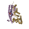
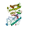
 Porphyromonas gingivalis (bacteria)
 X-RAY DIFFRACTION /
  Authors
 Citation
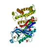
 Structure visualization
 Downloads & links
 Other downloads


 PDBj
PDBj Assembly
 Mass: 18.015 Da / Num. of mol.: 20 / Source method: isolated from a natural source / Formula: H2O
 Sample preparation
 / Beamline: X4C / Wavelength: 0.979 Å
 Processing