 Movie
Movie Controller


+ Open data

- Basic information
| Entry | Database: PDB / ID: 3d1d | ||||||
|---|---|---|---|---|---|---|---|
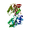
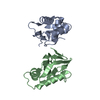
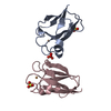
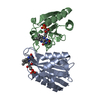
| Title | Hexagonal crystal structure of Tas3 C-terminal alpha motif | ||||||
 Components Components | RNA-induced transcriptional silencing complex protein tas3 | ||||||
 Keywords Keywords | NUCLEAR PROTEIN / all alpha motif / RITS complex immunoglobulin fold / Cell cycle / Chromosome partition / Nucleus / RNA-mediated gene silencing | ||||||
| Function / homology |  Function and homology information Function and homology informationRITS complex / subtelomeric heterochromatin / mating-type region heterochromatin / siRNA-mediated pericentric heterochromatin formation / pericentric heterochromatin formation / spindle pole body / siRNA binding / silent mating-type cassette heterochromatin formation / subtelomeric heterochromatin formation / pericentric heterochromatin ...RITS complex / subtelomeric heterochromatin / mating-type region heterochromatin / siRNA-mediated pericentric heterochromatin formation / pericentric heterochromatin formation / spindle pole body / siRNA binding / silent mating-type cassette heterochromatin formation / subtelomeric heterochromatin formation / pericentric heterochromatin / heterochromatin / chromosome segregation / heterochromatin formation / molecular adaptor activity / nucleus Similarity search - Function | ||||||
| Biological species |  | ||||||
| Method |  X-RAY DIFFRACTION / SYNCHROTRON / MOLECULAR REPLACEMENT / Resolution: 2.6 Å X-RAY DIFFRACTION / SYNCHROTRON / MOLECULAR REPLACEMENT / Resolution: 2.6 Å | ||||||
 Authors Authors | Li, H. / Patel, D.J. | ||||||
 Citation Citation | Journal: Mol.Cell / Year: 2009 Title: An alpha motif at Tas3 C terminus mediates RITS cis spreading and promotes heterochromatic gene silencing. Authors: Li, H. / Motamedi, M.R. / Yip, C.K. / Wang, Z. / Walz, T. / Patel, D.J. / Moazed, D. | ||||||
| History |
|
- Structure visualization
| Structure viewer | Molecule: MolmilJmol/JSmol |
|---|
- Downloads & links
-Download
| PDBx/mmCIF format | 3d1d.cif.gz | 140.1 KB | Display | PDBx/mmCIF format |
|---|---|---|---|---|
| PDB format | pdb3d1d.ent.gz | 112.2 KB | Display | PDB format |
| PDBx/mmJSON format | 3d1d.json.gz | Tree view | PDBx/mmJSON format | |
| Others |  Other downloads Other downloads |
-Validation report
| Arichive directory | https://data.pdbj.org/pub/pdb/validation_reports/d1/3d1dftp://data.pdbj.org/pub/pdb/validation_reports/d1/3d1d | HTTPS FTP |
|---|
-Related structure data
| Related structure data |  3d1bSC S: Starting model for refinement C: citing same article ( |
|---|---|
| Similar structure data |


-Links
 PDBj
PDBj- Assembly
| Deposited unit | 
| ||||||||
|---|---|---|---|---|---|---|---|---|---|
| 1 | 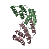
| ||||||||
| 2 | 
| ||||||||
| 3 | 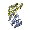
| ||||||||
| Unit cell |
|
-Components
| #1: Protein | Mass: 13940.937 Da / Num. of mol.: 6 / Fragment: UNP residues 426-545 Source method: isolated from a genetically manipulated source Source: (gene. exp.) Gene: tas3, SPBC83.03c / Production host:  #2: Water | ChemComp-HOH / |  Mass: 18.015 Da / Num. of mol.: 176 / Source method: isolated from a natural source / Formula: H2O Mass: 18.015 Da / Num. of mol.: 176 / Source method: isolated from a natural source / Formula: H2O |
|---|
-Experimental details
-Experiment
| Experiment | Method: X-RAY DIFFRACTION / Number of used crystals: 1 |
|---|
- Sample preparation
| Crystal | Density Matthews: 1.99 Å3/Da / Density % sol: 38.17 % |
|---|---|
| Crystal grow | Temperature: 293 K / Method: vapor diffusion, hanging drop / pH: 7.5 Details: 10% PEG4000, 0.1 M Tris 7.5, 0.2 M KCl, 50 mM MgCl2, and 15% ethylene glycol, VAPOR DIFFUSION, HANGING DROP, temperature 293K |
-Data collection
| Diffraction | Mean temperature: 100 K |
|---|---|
| Diffraction source | Source: SYNCHROTRON / Site: NSLS  / Beamline: X9A / Wavelength: 0.9791 Å / Beamline: X9A / Wavelength: 0.9791 Å |
| Detector | Type: MAR CCD 165 mm / Detector: CCD / Date: May 4, 2005 |
| Radiation | Protocol: SINGLE WAVELENGTH / Monochromatic (M) / Laue (L): M / Scattering type: x-ray |
| Radiation wavelength | Wavelength: 0.9791 Å / Relative weight: 1 |
| Reflection | Resolution: 2.6→20 Å / Num. all: 20741 / Num. obs: 19969 / % possible obs: 96.3 % / Observed criterion σ(F): -3 / Observed criterion σ(I): -3 / Redundancy: 12.2 % / Rmerge(I) obs: 0.109 / Net I/σ(I): 21.8 |
| Reflection shell | Resolution: 2.6→2.69 Å / Redundancy: 12 % / Rmerge(I) obs: 0.65 / Mean I/σ(I) obs: 2.6 / Num. unique all: 2052 / % possible all: 90 |
- Processing
| Software |
| |||||||||||||||||||||||||
|---|---|---|---|---|---|---|---|---|---|---|---|---|---|---|---|---|---|---|---|---|---|---|---|---|---|---|
| Refinement | Method to determine structure: MOLECULAR REPLACEMENT Starting model: PDB ENTRY 3D1B Resolution: 2.6→20 Å / Cross valid method: THROUGHOUT / σ(F): 0 / Stereochemistry target values: Engh & Huber
| |||||||||||||||||||||||||
| Refine analyze |
| |||||||||||||||||||||||||
| Refinement step | Cycle: LAST / Resolution: 2.6→20 Å
| |||||||||||||||||||||||||
| Refine LS restraints |
|
