 Movie
Movie Controller

[English] 日本語
 Yorodumi
- PDB-3cwk: Crystal Structure of the R132K:Y134F:R111L:T54V:L121E Mutant of C... -
+ Open data

- Basic information
| Entry | Database: PDB / ID: 3cwk | ||||||
|---|---|---|---|---|---|---|---|
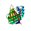
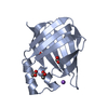
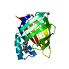
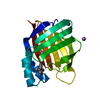
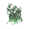
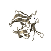
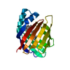
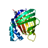
| Title | Crystal Structure of the R132K:Y134F:R111L:T54V:L121E Mutant of Cellular Retinoic Acid Binding Protein Type II in Complex with All-trans-Retinoic Acid at 1.57 Angstroms Resolution | ||||||
 Components Components | Cellular retinoic acid-binding protein 2 | ||||||
 Keywords Keywords | TRANSPORT PROTEIN / CRABPII / RETINOIC ACID / RETINOIDS / BETA BARREL / HIGH RESOLUTION / Cytoplasm / Nucleus / Retinol-binding / Transport / Vitamin A | ||||||
| Function / homology |  Function and homology information Function and homology informationpositive regulation of collateral sprouting / retinoid binding / retinoic acid binding / retinal binding / embryonic forelimb morphogenesis / retinoic acid metabolic process / retinol binding / Signaling by Retinoic Acid / epidermis development / fatty acid transport ...positive regulation of collateral sprouting / retinoid binding / retinoic acid binding / retinal binding / embryonic forelimb morphogenesis / retinoic acid metabolic process / retinol binding / Signaling by Retinoic Acid / epidermis development / fatty acid transport / cyclin binding / fatty acid binding / regulation of DNA-templated transcription / endoplasmic reticulum / signal transduction / extracellular exosome / nucleoplasm / nucleus / cytoplasm / cytosol Similarity search - Function | ||||||
| Biological species |  Homo sapiens (human) Homo sapiens (human) | ||||||
| Method |  X-RAY DIFFRACTION / SYNCHROTRON / RIGID BODY REFINEMENT / Resolution: 1.6 Å X-RAY DIFFRACTION / SYNCHROTRON / RIGID BODY REFINEMENT / Resolution: 1.6 Å | ||||||
 Authors Authors | Vaezeslami, S. / Geiger, J.H. | ||||||
 Citation Citation | Journal: Acta Crystallogr.,Sect.D / Year: 2008 Title: Structural analysis of site-directed mutants of cellular retinoic acid-binding protein II addresses the relationship between structural integrity and ligand binding. Authors: Vaezeslami, S. / Jia, X. / Vasileiou, C. / Borhan, B. / Geiger, J.H. #1: Journal: ThesisTitle: Determining Crystal Structures of Proteins and Protein Complexes by X-Ray Crystallography: X-Ray Crystallographic Studies of the Mutants of Cellular Retinoic Acid Binding Protein Type II ...Title: Determining Crystal Structures of Proteins and Protein Complexes by X-Ray Crystallography: X-Ray Crystallographic Studies of the Mutants of Cellular Retinoic Acid Binding Protein Type II Toward Designing a Mimic of Rhodopsin Authors: Vaezeslami, S. | ||||||
| History |
|
- Structure visualization
| Structure viewer | Molecule: MolmilJmol/JSmol |
|---|
- Downloads & links
-Download
| PDBx/mmCIF format | 3cwk.cif.gz | 80.5 KB | Display | PDBx/mmCIF format |
|---|---|---|---|---|
| PDB format | pdb3cwk.ent.gz | 61.3 KB | Display | PDB format |
| PDBx/mmJSON format | 3cwk.json.gz | Tree view | PDBx/mmJSON format | |
| Others |  Other downloads Other downloads |
-Validation report
| Arichive directory | https://data.pdbj.org/pub/pdb/validation_reports/cw/3cwkftp://data.pdbj.org/pub/pdb/validation_reports/cw/3cwk | HTTPS FTP |
|---|
-Related structure data


-Links
 PDBj
PDBj


- Assembly
| Deposited unit | 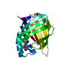
| ||||||||
|---|---|---|---|---|---|---|---|---|---|
| 1 |
| ||||||||
| Unit cell |
|
-Components
| #1: Protein | Mass: 15507.735 Da / Num. of mol.: 1 / Mutation: T54V, R111L, L121E, R132K, Y134F Source method: isolated from a genetically manipulated source Source: (gene. exp.) Homo sapiens (human) / Gene: CRABP2 / Plasmid: PET17-B / Production host:  |
|---|---|
| #2: Chemical | ChemComp-SO4 /   Mass: 96.063 Da / Num. of mol.: 1 / Source method: obtained synthetically / Formula: SO4 Mass: 96.063 Da / Num. of mol.: 1 / Source method: obtained synthetically / Formula: SO4 |
| #3: Chemical | ChemComp-REA /   Mass: 300.435 Da / Num. of mol.: 1 / Source method: obtained synthetically / Formula: C20H28O2 Mass: 300.435 Da / Num. of mol.: 1 / Source method: obtained synthetically / Formula: C20H28O2 |
| #4: Water | ChemComp-HOH /  Mass: 18.015 Da / Num. of mol.: 221 / Source method: isolated from a natural source / Formula: H2O Mass: 18.015 Da / Num. of mol.: 221 / Source method: isolated from a natural source / Formula: H2O |
-Experimental details
-Experiment
| Experiment | Method: X-RAY DIFFRACTION / Number of used crystals: 1 |
|---|
- Sample preparation
| Crystal | Density Matthews: 2.58 Å3/Da / Density % sol: 52.29 % |
|---|---|
| Crystal grow | Temperature: 298 K / Method: vapor diffusion, hanging drop / pH: 6.5 Details: 0.1 M MES, 30% (W/V) PEG 5000, 0.2 M AMMONIUM SULFATE, pH 6.50, VAPOR DIFFUSION, HANGING DROP, temperature 298K |
-Data collection
| Diffraction | Mean temperature: 77 K |
|---|---|
| Diffraction source | Source: SYNCHROTRON / Site: APS  / Beamline: 32-ID / Wavelength: 1 / Beamline: 32-ID / Wavelength: 1 |
| Detector | Type: MARRESEARCH / Detector: CCD / Date: Mar 13, 2004 |
| Radiation | Protocol: SINGLE WAVELENGTH / Monochromatic (M) / Laue (L): M / Scattering type: x-ray |
| Radiation wavelength | Wavelength: 1 Å / Relative weight: 1 |
| Reflection | Resolution: 1.6→50 Å / Num. obs: 21826 / % possible obs: 99.8 % / Observed criterion σ(I): -3 / Rmerge(I) obs: 0.05 / Net I/σ(I): 27.28 |
| Reflection shell | Resolution: 1.6→1.66 Å / Rmerge(I) obs: 0.157 / % possible all: 100 |
- Processing
| Software |
| ||||||||||||||||||||||||||||||||||||||||||||||||||||||||||||||||||||||||||||||||||||||||||||||||||||||||||||||||||||||||||||||||||||||||||||||||||||||||||||||||||||||||||
|---|---|---|---|---|---|---|---|---|---|---|---|---|---|---|---|---|---|---|---|---|---|---|---|---|---|---|---|---|---|---|---|---|---|---|---|---|---|---|---|---|---|---|---|---|---|---|---|---|---|---|---|---|---|---|---|---|---|---|---|---|---|---|---|---|---|---|---|---|---|---|---|---|---|---|---|---|---|---|---|---|---|---|---|---|---|---|---|---|---|---|---|---|---|---|---|---|---|---|---|---|---|---|---|---|---|---|---|---|---|---|---|---|---|---|---|---|---|---|---|---|---|---|---|---|---|---|---|---|---|---|---|---|---|---|---|---|---|---|---|---|---|---|---|---|---|---|---|---|---|---|---|---|---|---|---|---|---|---|---|---|---|---|---|---|---|---|---|---|---|---|---|
| Refinement | Method to determine structure: RIGID BODY REFINEMENT / Resolution: 1.6→39.47 Å / Cor.coef. Fo:Fc: 0.975 / Cor.coef. Fo:Fc free: 0.96 / SU B: 2.15 / SU ML: 0.036 / Cross valid method: THROUGHOUT / ESU R: 0.093 / ESU R Free: 0.076 / Stereochemistry target values: MAXIMUM LIKELIHOOD / Details: HYDROGENS HAVE BEEN ADDED IN THE RIDING POSITIONS
| ||||||||||||||||||||||||||||||||||||||||||||||||||||||||||||||||||||||||||||||||||||||||||||||||||||||||||||||||||||||||||||||||||||||||||||||||||||||||||||||||||||||||||
| Solvent computation | Ion probe radii: 0.8 Å / Shrinkage radii: 0.8 Å / VDW probe radii: 1.2 Å / Solvent model: BABINET MODEL WITH MASK | ||||||||||||||||||||||||||||||||||||||||||||||||||||||||||||||||||||||||||||||||||||||||||||||||||||||||||||||||||||||||||||||||||||||||||||||||||||||||||||||||||||||||||
| Displacement parameters | Biso mean: 14.29 Å2
| ||||||||||||||||||||||||||||||||||||||||||||||||||||||||||||||||||||||||||||||||||||||||||||||||||||||||||||||||||||||||||||||||||||||||||||||||||||||||||||||||||||||||||
| Refinement step | Cycle: LAST / Resolution: 1.6→39.47 Å
| ||||||||||||||||||||||||||||||||||||||||||||||||||||||||||||||||||||||||||||||||||||||||||||||||||||||||||||||||||||||||||||||||||||||||||||||||||||||||||||||||||||||||||
| Refine LS restraints |
| ||||||||||||||||||||||||||||||||||||||||||||||||||||||||||||||||||||||||||||||||||||||||||||||||||||||||||||||||||||||||||||||||||||||||||||||||||||||||||||||||||||||||||
| LS refinement shell | Resolution: 1.6→1.64 Å / Total num. of bins used: 20
|
