 Movie
Movie Controller


+ Open data

- Basic information
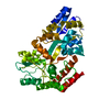
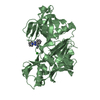
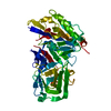
| Entry | Database: PDB / ID: 3cv3 | ||||||
|---|---|---|---|---|---|---|---|
| Title | Crystal Structure of GumK mutant D157A in complex with UDP | ||||||
 Components Components | Glucuronosyltransferase GumK | ||||||
 Keywords Keywords | TRANSFERASE / Glucuronosyltransferase / glycosyltransferase / xanthan / Xanthomonas campestris / UDP / UDPGlcA | ||||||
| Function / homology |  Function and homology information Function and homology informationD-Man-alpha-(1->3)-D-Glc-beta-(1->4)-D-Glc-alpha-1-diphosphoundecaprenol 2-beta-glucuronosyltransferase / glucuronosyltransferase activity / polysaccharide biosynthetic process / plasma membrane Similarity search - Function | ||||||
| Biological species |  Xanthomonas campestris pv. campestris (bacteria) Xanthomonas campestris pv. campestris (bacteria) | ||||||
| Method |  X-RAY DIFFRACTION / SYNCHROTRON / MOLECULAR REPLACEMENT / molecular replacement / Resolution: 2.25 Å X-RAY DIFFRACTION / SYNCHROTRON / MOLECULAR REPLACEMENT / molecular replacement / Resolution: 2.25 Å | ||||||
 Authors Authors | Barreras, M. | ||||||
 Citation Citation | Journal: J.Biol.Chem. / Year: 2008 Title: Structure and mechanism of GumK, a membrane-associated glucuronosyltransferase. Authors: Barreras, M. / Salinas, S.R. / Abdian, P.L. / Kampel, M.A. / Ielpi, L. | ||||||
| History |
|
- Structure visualization
| Structure viewer | Molecule: MolmilJmol/JSmol |
|---|
- Downloads & links
-Download
| PDBx/mmCIF format | 3cv3.cif.gz | 99.1 KB | Display | PDBx/mmCIF format |
|---|---|---|---|---|
| PDB format | pdb3cv3.ent.gz | 73.5 KB | Display | PDB format |
| PDBx/mmJSON format | 3cv3.json.gz | Tree view | PDBx/mmJSON format | |
| Others |  Other downloads Other downloads |
-Validation report
| Arichive directory | https://data.pdbj.org/pub/pdb/validation_reports/cv/3cv3ftp://data.pdbj.org/pub/pdb/validation_reports/cv/3cv3 | HTTPS FTP |
|---|
-Related structure data
| Related structure data | 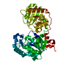 2hy7SC 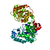 2q6vC 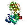 3cuyC C: citing same article ( S: Starting model for refinement |
|---|---|
| Similar structure data |


-Links
 PDBj
PDBj


- Assembly
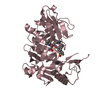
| Deposited unit | 
| ||||||||||||
|---|---|---|---|---|---|---|---|---|---|---|---|---|---|
| 1 |
| ||||||||||||
| Unit cell |
| ||||||||||||
| Components on special symmetry positions |
|
-Components
| #1: Protein | Mass: 45291.426 Da / Num. of mol.: 1 / Mutation: D157A Source method: isolated from a genetically manipulated source Source: (gene. exp.) Xanthomonas campestris pv. campestris (bacteria)Gene: gumK / Plasmid: pET22b / Production host: |
|---|---|
| #2: Chemical | ChemComp-UDP /   Type: RNA linking / Mass: 404.161 Da / Num. of mol.: 1 / Source method: obtained synthetically / Formula: C9H14N2O12P2 / Comment: UDP*YM Type: RNA linking / Mass: 404.161 Da / Num. of mol.: 1 / Source method: obtained synthetically / Formula: C9H14N2O12P2 / Comment: UDP*YM |
| #3: Water | ChemComp-HOH /  Mass: 18.015 Da / Num. of mol.: 422 / Source method: isolated from a natural source / Formula: H2O Mass: 18.015 Da / Num. of mol.: 422 / Source method: isolated from a natural source / Formula: H2O |
-Experimental details
-Experiment
| Experiment | Method: X-RAY DIFFRACTION / Number of used crystals: 1 |
|---|
- Sample preparation
| Crystal | Density Matthews: 3.99 Å3/Da / Density % sol: 69.15 % |
|---|---|
| Crystal grow | Temperature: 293 K / Method: vapor diffusion, hanging drop / pH: 8.2 Details: 0.1 M Tris-HCl, 35% (w/v) PEG 3350, 0.2 M Li2SO4, 0.1 M LiCl, pH 8.2, hanging drop and soaking of crystals in UDPGlcA 10 mM, temperature 293K, VAPOR DIFFUSION, HANGING DROP |
-Data collection
| Diffraction | Mean temperature: 100 K |
|---|---|
| Diffraction source | Source: SYNCHROTRON / Site: LNLS  / Beamline: D03B-MX1 / Wavelength: 1.433 Å / Beamline: D03B-MX1 / Wavelength: 1.433 Å |
| Detector | Type: MARRESEARCH / Detector: CCD / Date: Apr 1, 2008 / Details: beam focused by a cilindrically curved mirror |
| Radiation | Monochromator: Silicium curved crystal, with asymmetric 7.25 angle cut Protocol: SINGLE WAVELENGTH / Monochromatic (M) / Laue (L): M / Scattering type: x-ray |
| Radiation wavelength | Wavelength: 1.433 Å / Relative weight: 1 |
| Reflection | Resolution: 2.25→17.46 Å / Num. obs: 35644 / % possible obs: 99.8 % / Observed criterion σ(F): 1 / Observed criterion σ(I): 1 / Redundancy: 14.9 % / Biso Wilson estimate: 25.9 Å2 / Rmerge(I) obs: 0.149 / Rsym value: 0.149 / Net I/σ(I): 16.8 |
| Reflection shell | Resolution: 2.25→2.37 Å / Redundancy: 14.5 % / Rmerge(I) obs: 0.286 / Mean I/σ(I) obs: 10.1 / Num. unique all: 5131 / Rsym value: 0.297 / % possible all: 100 |
-Phasing
| Phasing | Method: molecular replacement |
|---|
- Processing
| Software |
| ||||||||||||||||||||||||||||||||||||||||||||||||||||||||||||||||||||||||||||||||||||||||||
|---|---|---|---|---|---|---|---|---|---|---|---|---|---|---|---|---|---|---|---|---|---|---|---|---|---|---|---|---|---|---|---|---|---|---|---|---|---|---|---|---|---|---|---|---|---|---|---|---|---|---|---|---|---|---|---|---|---|---|---|---|---|---|---|---|---|---|---|---|---|---|---|---|---|---|---|---|---|---|---|---|---|---|---|---|---|---|---|---|---|---|---|
| Refinement | Method to determine structure: MOLECULAR REPLACEMENT Starting model: PDB entry 2HY7 Resolution: 2.25→17.46 Å / Cor.coef. Fo:Fc: 0.935 / Cor.coef. Fo:Fc free: 0.894 / SU B: 3.927 / SU ML: 0.1 / Cross valid method: THROUGHOUT / σ(F): 0 / ESU R: 0.161 / ESU R Free: 0.157 / Stereochemistry target values: MAXIMUM LIKELIHOOD
| ||||||||||||||||||||||||||||||||||||||||||||||||||||||||||||||||||||||||||||||||||||||||||
| Solvent computation | Ion probe radii: 0.8 Å / Shrinkage radii: 0.8 Å / VDW probe radii: 1.2 Å / Solvent model: MASK | ||||||||||||||||||||||||||||||||||||||||||||||||||||||||||||||||||||||||||||||||||||||||||
| Displacement parameters | Biso mean: 15.864 Å2
| ||||||||||||||||||||||||||||||||||||||||||||||||||||||||||||||||||||||||||||||||||||||||||
| Refinement step | Cycle: LAST / Resolution: 2.25→17.46 Å
| ||||||||||||||||||||||||||||||||||||||||||||||||||||||||||||||||||||||||||||||||||||||||||
| Refine LS restraints |
| ||||||||||||||||||||||||||||||||||||||||||||||||||||||||||||||||||||||||||||||||||||||||||
| LS refinement shell | Resolution: 2.25→2.307 Å / Total num. of bins used: 20
|
