Mass: 18.015 Da / Num. of mol.: 95 / Source method: isolated from a natural source / Formula: H2O
-
Details
Has protein modification
Y
Sequence details
THE CONSTRUCT WAS EXPRESSED WITH A PURIFICATION TAG MGSDKIHHHHHHENLYFQG. THE TAG WAS REMOVED WITH ...THE CONSTRUCT WAS EXPRESSED WITH A PURIFICATION TAG MGSDKIHHHHHHENLYFQG. THE TAG WAS REMOVED WITH TEV PROTEASE LEAVING ONLY A GLYCINE FOLLOWED BY THE TARGET SEQUENCE.
-
Experimental details
-
Experiment
Experiment
Method: X-RAY DIFFRACTION / Number of used crystals: 1
-
Sample preparation
Crystal
Density Matthews: 2.67 Å3/Da / Density % sol: 53.93 %
Crystal grow
Temperature: 293 K / Method: vapor diffusion, sitting drop / pH: 7.04 Details: NANODROP, 34.5% PEG 400, 0.2M Magnesium chloride, 0.1M HEPES pH 7.04, VAPOR DIFFUSION, SITTING DROP, temperature 293K
Resolution: 2.1→29.553 Å / Num. obs: 35037 / % possible obs: 97.4 % / Observed criterion σ(I): -3 / Biso Wilson estimate: 36.671 Å2 / Rmerge(I) obs: 0.039 / Net I/σ(I): 13.7
Reflection shell
Resolution (Å)
Rmerge(I) obs
Mean I/σ(I) obs
Num. measured obs
Num. unique obs
Diffraction-ID
% possible all
2.1-2.17
0.458
1.9
10175
5897
1
94.9
2.17-2.26
0.373
2.5
11857
6779
1
97.8
2.26-2.36
0.285
3.1
11115
6391
1
98
2.36-2.49
0.224
3.9
12029
6842
1
97.7
2.49-2.64
0.166
5.2
11106
6308
1
97.7
2.64-2.85
0.107
7.9
11874
6721
1
98
2.85-3.13
0.065
12.2
11372
6401
1
97.8
3.13-3.59
0.032
22.5
11823
6663
1
98.1
3.59-4.51
0.019
34.6
11496
6480
1
98
4.51-29.553
0.014
42.8
11642
6445
1
95.6
-
Phasing
Phasing
Method: MAD
-
Processing
Software
Name
Version
Classification
NB
REFMAC
5.4.0067
refinement
PHENIX
refinement
SHELX
phasing
MolProbity
3beta29
modelbuilding
XSCALE
datascaling
PDB_EXTRACT
3.004
dataextraction
MAR345
CCD
datacollection
XDS
datareduction
SHARP
phasing
SHELXD
phasing
Refinement
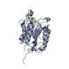
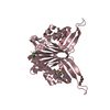
Method to determine structure: MAD / Resolution: 2.1→29.553 Å / Cor.coef. Fo:Fc: 0.953 / Cor.coef. Fo:Fc free: 0.937 / SU B: 9.083 / SU ML: 0.121 / TLS residual ADP flag: LIKELY RESIDUAL / Cross valid method: THROUGHOUT / σ(F): 0 / ESU R: 0.179 / ESU R Free: 0.162 Stereochemistry target values: MAXIMUM LIKELIHOOD WITH PHASES Details: 1. HYDROGENS HAVE BEEN ADDED IN THE RIDING POSITIONS. 2. ATOM RECORD CONTAINS RESIDUAL B FACTORS ONLY. 3. A MET-INHIBITION PROTOCOL WAS USED FOR SELENOMETHIONINE INCORPORATION DURING PROTEIN ...Details: 1. HYDROGENS HAVE BEEN ADDED IN THE RIDING POSITIONS. 2. ATOM RECORD CONTAINS RESIDUAL B FACTORS ONLY. 3. A MET-INHIBITION PROTOCOL WAS USED FOR SELENOMETHIONINE INCORPORATION DURING PROTEIN EXPRESSION. THE OCCUPANCY OF THE SE ATOMS IN THE MSE RESIDUES WAS REDUCED TO 0.75 FOR THE REDUCED SCATTERING POWER DUE TO PARTIAL S-MET INCORPORATION 3. CHLORIDE, MAGNESIUM, PARTIAL PEG(S) AND GLYCEROL WERE MODELED BASED ON CRYSTALLIZATION AND CRYOPROTECTION CONDITIONS. 4. AN UNIDENTIFIED LIGAND (UNL) HAS BEEN TENTATIVELY MODELED AT WHAT APPEARS TO BE PUTATIVE ACTIVE SITE(S) WHICH COULD BE A PROTEASE-LIKE CATALYTIC TRIAD FORMED BY CYS59-HIS150-E109 OR A METAL BINDING SITE FORMED BY HIS61-HIS150-E109.
Rfactor
Num. reflection
% reflection
Selection details
Rfree
0.235
1752
5 %
RANDOM
Rwork
0.199
-
-
-
obs
0.201
34988
99.03 %
-
Solvent computation
Ion probe radii: 0.8 Å / Shrinkage radii: 0.8 Å / VDW probe radii: 1.2 Å / Solvent model: MASK
Displacement parameters
Biso mean: 34.351 Å2
Baniso -1
Baniso -2
Baniso -3
1-
1.21 Å2
0 Å2
0 Å2
2-
-
0.8 Å2
0 Å2
3-
-
-
-2.02 Å2
Refinement step
Cycle: LAST / Resolution: 2.1→29.553 Å
Protein
Nucleic acid
Ligand
Solvent
Total
Num. atoms
3221
0
88
95
3404
Refine LS restraints
Refine-ID
Type
Dev ideal
Dev ideal target
Number
X-RAY DIFFRACTION
r_bond_refined_d
0.016
0.022
3409
X-RAY DIFFRACTION
r_bond_other_d
0.001
0.02
2198
X-RAY DIFFRACTION
r_angle_refined_deg
1.668
1.96
4655
X-RAY DIFFRACTION
r_angle_other_deg
1.447
3
5387
X-RAY DIFFRACTION
r_dihedral_angle_1_deg
6.071
5
437
X-RAY DIFFRACTION
r_dihedral_angle_2_deg
34.9
23.876
129
X-RAY DIFFRACTION
r_dihedral_angle_3_deg
15.587
15
465
X-RAY DIFFRACTION
r_dihedral_angle_4_deg
15.308
15
15
X-RAY DIFFRACTION
r_chiral_restr
0.083
0.2
543
X-RAY DIFFRACTION
r_gen_planes_refined
0.008
0.021
3753
X-RAY DIFFRACTION
r_gen_planes_other
0.002
0.02
662
X-RAY DIFFRACTION
r_mcbond_it
0.731
1.5
2162
X-RAY DIFFRACTION
r_mcbond_other
0.19
1.5
879
X-RAY DIFFRACTION
r_mcangle_it
1.321
2
3490
X-RAY DIFFRACTION
r_scbond_it
2.099
3
1247
X-RAY DIFFRACTION
r_scangle_it
2.905
4.5
1159
Refine LS restraints NCS
Ens-ID: 1 / Refine-ID: X-RAY DIFFRACTION
Dom-ID
Auth asym-ID
Number
Type
Rms dev position (Å)
Weight position
1
A
786
MEDIUMPOSITIONAL
0.29
0.5
2
B
786
MEDIUMPOSITIONAL
0.19
0.5
3
C
786
MEDIUMPOSITIONAL
0.21
0.5
1
A
839
LOOSEPOSITIONAL
0.41
5
2
B
839
LOOSEPOSITIONAL
0.38
5
3
C
839
LOOSEPOSITIONAL
0.38
5
1
A
786
MEDIUMTHERMAL
0.68
2
2
B
786
MEDIUMTHERMAL
0.54
2
3
C
786
MEDIUMTHERMAL
0.59
2
1
A
839
LOOSETHERMAL
0.82
10
2
B
839
LOOSETHERMAL
0.87
10
3
C
839
LOOSETHERMAL
0.92
10
LS refinement shell
Resolution: 2.1→2.154 Å / Total num. of bins used: 20
Rfactor
Num. reflection
% reflection
Rfree
0.279
116
-
Rwork
0.247
2369
-
all
-
2485
-
obs
-
-
97.49 %
Refinement TLS params.
Method: refined / Refine-ID: X-RAY DIFFRACTION
ID
L11 (°2)
L12 (°2)
L13 (°2)
L22 (°2)
L23 (°2)
L33 (°2)
S11 (Å °)
S12 (Å °)
S13 (Å °)
S21 (Å °)
S22 (Å °)
S23 (Å °)
S31 (Å °)
S32 (Å °)
S33 (Å °)
T11 (Å2)
T12 (Å2)
T13 (Å2)
T22 (Å2)
T23 (Å2)
T33 (Å2)
Origin x (Å)
Origin y (Å)
Origin z (Å)
1
3.0149
1.29
-0.66
2.6928
-0.7505
3.0703
0.0723
-0.2359
-0.2915
0.3037
-0.2958
-0.1462
0.1534
0.2614
0.2234
-0.012
-0.1317
-0.0379
-0.0758
0.0375
-0.1223
86.214
15.1585
34.6524
2
1.3017
0.8973
-0.1049
2.5264
-1.0674
2.2755
-0.0945
0.1642
-0.1187
-0.1861
-0.1703
-0.2611
0.1955
0.3263
0.2648
-0.085
-0.0989
-0.011
0.0041
0.0344
-0.0979
93.7755
24.6218
15.6337
3
1.8411
0.2798
0.5329
1.7911
-0.6619
1.3475
0.1711
0.1037
-0.1947
-0.2359
-0.065
-0.0624
0.2422
0.0092
-0.1061
-0.0663
0.0161
-0.037
-0.1104
-0.0209
-0.0794
105.2487
52.7529
2.8696
Refinement TLS group
ID
Refine-ID
Refine TLS-ID
Auth asym-ID
Label asym-ID
Auth seq-ID
Label seq-ID
1
X-RAY DIFFRACTION
1
A
A
7 - 157
8 - 158
2
X-RAY DIFFRACTION
2
B
B
7 - 157
8 - 158
3
X-RAY DIFFRACTION
3
C
C
6 - 156
7 - 157
+
About Yorodumi
-
News
-
Feb 9, 2022. New format data for meta-information of EMDB entries
New format data for meta-information of EMDB entries
Version 3 of the EMDB header file is now the official format.
The previous official version 1.9 will be removed from the archive.
In the structure databanks used in Yorodumi, some data are registered as the other names, "COVID-19 virus" and "2019-nCoV". Here are the details of the virus and the list of structure data.
Jan 31, 2019. EMDB accession codes are about to change! (news from PDBe EMDB page)
EMDB accession codes are about to change! (news from PDBe EMDB page)
The allocation of 4 digits for EMDB accession codes will soon come to an end. Whilst these codes will remain in use, new EMDB accession codes will include an additional digit and will expand incrementally as the available range of codes is exhausted. The current 4-digit format prefixed with “EMD-” (i.e. EMD-XXXX) will advance to a 5-digit format (i.e. EMD-XXXXX), and so on. It is currently estimated that the 4-digit codes will be depleted around Spring 2019, at which point the 5-digit format will come into force.
The EM Navigator/Yorodumi systems omit the EMD- prefix.
Related info.:Q: What is EMD? / ID/Accession-code notation in Yorodumi/EM Navigator
Yorodumi is a browser for structure data from EMDB, PDB, SASBDB, etc.
This page is also the successor to EM Navigator detail page, and also detail information page/front-end page for Omokage search.
The word "yorodu" (or yorozu) is an old Japanese word meaning "ten thousand". "mi" (miru) is to see.
Related info.:EMDB / PDB / SASBDB / Comparison of 3 databanks / Yorodumi Search / Aug 31, 2016. New EM Navigator & Yorodumi / Yorodumi Papers / Jmol/JSmol / Function and homology information / Changes in new EM Navigator and Yorodumi
 Movie
Movie Controller

 Yorodumi
 Open data
 Basic information
 Components
 Keywords
 Function and homology information
 Streptomyces avermitilis (bacteria)
 X-RAY DIFFRACTION /
  Authors
 Citation
 Structure visualization
 Downloads & links
 Other downloads

 PDBj
PDBj


 Assembly
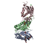


 Mass: 35.453 Da / Num. of mol.: 3 / Source method: obtained synthetically / Formula: Cl
 Mass: 150.173 Da / Num. of mol.: 1 / Source method: obtained synthetically / Formula: C6H14O4
 Mass: 106.120 Da / Num. of mol.: 4 / Source method: obtained synthetically / Formula: C4H10O3
 Mass: 24.305 Da / Num. of mol.: 2 / Source method: obtained synthetically / Formula: Mg
 Mass: 266.331 Da / Num. of mol.: 1 / Source method: obtained synthetically / Formula: C12H26O6
 Mass: 92.094 Da / Num. of mol.: 3 / Source method: obtained synthetically / Formula: C3H8O3
 Sample preparation
 / Beamline: BL9-2 / Wavelength: 0.91162, 0.97978
 Processing