Mass: 18.015 Da / Num. of mol.: 323 / Source method: isolated from a natural source / Formula: H2O
-
Details
Has protein modification
Y
Sequence details
THE CONSTRUCT WAS EXPRESSED WITH A PURIFICATION TAG MGSDKIHHHHHHENLYFQG. THE TAG WAS REMOVED WITH ...THE CONSTRUCT WAS EXPRESSED WITH A PURIFICATION TAG MGSDKIHHHHHHENLYFQG. THE TAG WAS REMOVED WITH TEV PROTEASE LEAVING ONLY A GLYCINE (0) FOLLOWED BY THE TARGET SEQUENCE.
-
Experimental details
-
Experiment
Experiment
Method: X-RAY DIFFRACTION / Number of used crystals: 1
-
Sample preparation
Crystal
Density Matthews: 2.38 Å3/Da / Density % sol: 48.4 %
Crystal grow
Temperature: 277 K / Method: vapor diffusion, sitting drop / pH: 4.2 Details: NANODROP, 0.2M NaCl, 20.0% PEG 8000, 0.1M Phosphate citrate pH 4.2, VAPOR DIFFUSION, SITTING DROP, temperature 277K
Resolution: 1.66→35.355 Å / Num. obs: 53836 / % possible obs: 99.8 % / Observed criterion σ(I): -3 / Biso Wilson estimate: 24.504 Å2 / Rmerge(I) obs: 0.055 / Net I/σ(I): 17.74
Reflection shell
Resolution (Å)
Rmerge(I) obs
Mean I/σ(I) obs
Num. measured obs
Num. unique obs
Diffraction-ID
% possible all
1.66-1.72
0.716
2.1
27325
5323
1
99.7
1.72-1.79
0.514
3.4
36734
5372
1
99.9
1.79-1.87
0.335
5.2
36685
5205
1
100
1.87-1.97
0.242
7.6
38301
5385
1
100
1.97-2.09
0.152
11.6
37321
5189
1
100
2.09-2.25
0.102
17
38506
5344
1
100
2.25-2.48
0.073
22.4
39259
5467
1
100
2.48-2.84
0.058
27.9
38702
5429
1
100
2.84-3.57
0.039
35.8
38164
5439
1
99.9
3.57-35.355
0.031
41.5
38189
5780
1
99
-
Phasing
Phasing
Method: MAD
-
Processing
Software
Name
Version
Classification
NB
REFMAC
5.4.0067
refinement
PHENIX
refinement
SHELX
phasing
MolProbity
3beta29
modelbuilding
XSCALE
datascaling
PDB_EXTRACT
3
dataextraction
MAR345
CCD
datacollection
XDS
datareduction
SHELXD
phasing
autoSHARP
phasing
Refinement
Method to determine structure: MAD / Resolution: 1.66→35.355 Å / Cor.coef. Fo:Fc: 0.971 / Cor.coef. Fo:Fc free: 0.963 / SU B: 3.601 / SU ML: 0.06 / TLS residual ADP flag: LIKELY RESIDUAL / Cross valid method: THROUGHOUT / σ(F): 0 / ESU R: 0.088 / ESU R Free: 0.086 Stereochemistry target values: MAXIMUM LIKELIHOOD WITH PHASES Details: 1. HYDROGENS HAVE BEEN ADDED IN THE RIDING POSITIONS. 2. ATOM RECORDS CONTAIN RESIDUAL B FACTORS ONLY. 3. A MET-INHIBITION PROTOCOL WAS USED FOR SELENOMETHIONINE INCORPORATION DURING PROTEIN ...Details: 1. HYDROGENS HAVE BEEN ADDED IN THE RIDING POSITIONS. 2. ATOM RECORDS CONTAIN RESIDUAL B FACTORS ONLY. 3. A MET-INHIBITION PROTOCOL WAS USED FOR SELENOMETHIONINE INCORPORATION DURING PROTEIN EXPRESSION. THE OCCUPANCY OF THE SE ATOMS IN THE MSE RESIDUES WAS REDUCED TO 0.75 FOR THE REDUCED SCATTERING POWER DUE TO PARTIAL S-MET INCORPORATION. 4. NA ION, GLYCEROL (GOL), PHOSPHATE ION (PO4) AND PEG-8000 (PG4) MOLECULES FROM THE CRYSTALLIZATION/CRYO SOLUTION ARE MODELED. 5. THE OCCUPANCY OF PO4 AND GOL HAS BEEN REDUCED FOR OPTIMAL REFINEMENT.
Rfactor
Num. reflection
% reflection
Selection details
Rfree
0.187
2734
5.1 %
RANDOM
Rwork
0.16
-
-
-
obs
0.161
53757
99.87 %
-
Solvent computation
Ion probe radii: 0.8 Å / Shrinkage radii: 0.8 Å / VDW probe radii: 1.2 Å / Solvent model: BABINET MODEL WITH MASK
Displacement parameters
Biso mean: 29.653 Å2
Baniso -1
Baniso -2
Baniso -3
1-
-0.76 Å2
0 Å2
0 Å2
2-
-
-0.76 Å2
0 Å2
3-
-
-
1.53 Å2
Refinement step
Cycle: LAST / Resolution: 1.66→35.355 Å
Protein
Nucleic acid
Ligand
Solvent
Total
Num. atoms
3175
0
28
323
3526
Refine LS restraints
Refine-ID
Type
Dev ideal
Dev ideal target
Number
X-RAY DIFFRACTION
r_bond_refined_d
0.016
0.022
3544
X-RAY DIFFRACTION
r_bond_other_d
0.002
0.02
2537
X-RAY DIFFRACTION
r_angle_refined_deg
1.669
1.974
4841
X-RAY DIFFRACTION
r_angle_other_deg
1.805
3
6108
X-RAY DIFFRACTION
r_dihedral_angle_1_deg
4.467
5
467
X-RAY DIFFRACTION
r_dihedral_angle_2_deg
30.043
22.438
201
X-RAY DIFFRACTION
r_dihedral_angle_3_deg
11.921
15
632
X-RAY DIFFRACTION
r_dihedral_angle_4_deg
11.824
15
56
X-RAY DIFFRACTION
r_chiral_restr
0.102
0.2
523
X-RAY DIFFRACTION
r_gen_planes_refined
0.008
0.02
4076
X-RAY DIFFRACTION
r_gen_planes_other
0.003
0.02
820
X-RAY DIFFRACTION
r_mcbond_it
1.095
2
2104
X-RAY DIFFRACTION
r_mcbond_other
0.234
2
846
X-RAY DIFFRACTION
r_mcangle_it
2.019
4
3408
X-RAY DIFFRACTION
r_scbond_it
3.623
6
1440
X-RAY DIFFRACTION
r_scangle_it
5.774
8
1395
LS refinement shell
Resolution: 1.66→1.703 Å / Total num. of bins used: 20
Rfactor
Num. reflection
% reflection
Rfree
0.273
221
-
Rwork
0.228
3668
-
all
-
3889
-
obs
-
-
99.74 %
Refinement TLS params.
Method: refined / Refine-ID: X-RAY DIFFRACTION
ID
L11 (°2)
L12 (°2)
L13 (°2)
L22 (°2)
L23 (°2)
L33 (°2)
S11 (Å °)
S12 (Å °)
S13 (Å °)
S21 (Å °)
S22 (Å °)
S23 (Å °)
S31 (Å °)
S32 (Å °)
S33 (Å °)
T11 (Å2)
T12 (Å2)
T13 (Å2)
T22 (Å2)
T23 (Å2)
T33 (Å2)
Origin x (Å)
Origin y (Å)
Origin z (Å)
1
0.7413
0.0856
-0.2939
0.7202
-0.134
1.1873
0.0123
0.0898
0.0237
-0.0374
0.0259
0.0569
-0.0363
-0.1281
-0.0382
-0.1089
-0.0011
-0.0154
-0.0857
-0.001
-0.2738
67.367
72.493
107.318
2
1.5771
-0.2419
0.1873
0.4151
0.0399
0.7298
0.009
0.1521
-0.1532
-0.0177
-0.0335
-0.0274
0.0328
0.072
0.0245
-0.0837
0.0039
0.0054
-0.0897
-0.0204
-0.228
92.181
54.815
106.295
Refinement TLS group
ID
Refine-ID
Refine TLS-ID
Auth asym-ID
Label asym-ID
Auth seq-ID
Label seq-ID
1
X-RAY DIFFRACTION
1
A
A
0 - 199
1 - 200
2
X-RAY DIFFRACTION
2
B
B
1 - 199
2 - 200
+
About Yorodumi
-
News
-
Feb 9, 2022. New format data for meta-information of EMDB entries
New format data for meta-information of EMDB entries
Version 3 of the EMDB header file is now the official format.
The previous official version 1.9 will be removed from the archive.
In the structure databanks used in Yorodumi, some data are registered as the other names, "COVID-19 virus" and "2019-nCoV". Here are the details of the virus and the list of structure data.
Jan 31, 2019. EMDB accession codes are about to change! (news from PDBe EMDB page)
EMDB accession codes are about to change! (news from PDBe EMDB page)
The allocation of 4 digits for EMDB accession codes will soon come to an end. Whilst these codes will remain in use, new EMDB accession codes will include an additional digit and will expand incrementally as the available range of codes is exhausted. The current 4-digit format prefixed with “EMD-” (i.e. EMD-XXXX) will advance to a 5-digit format (i.e. EMD-XXXXX), and so on. It is currently estimated that the 4-digit codes will be depleted around Spring 2019, at which point the 5-digit format will come into force.
The EM Navigator/Yorodumi systems omit the EMD- prefix.
Related info.:Q: What is EMD? / ID/Accession-code notation in Yorodumi/EM Navigator
Yorodumi is a browser for structure data from EMDB, PDB, SASBDB, etc.
This page is also the successor to EM Navigator detail page, and also detail information page/front-end page for Omokage search.
The word "yorodu" (or yorozu) is an old Japanese word meaning "ten thousand". "mi" (miru) is to see.
Related info.:EMDB / PDB / SASBDB / Comparison of 3 databanks / Yorodumi Search / Aug 31, 2016. New EM Navigator & Yorodumi / Yorodumi Papers / Jmol/JSmol / Function and homology information / Changes in new EM Navigator and Yorodumi
 Movie
Movie Controller

 Yorodumi
 Open data
 Basic information
 Components
 Keywords
 Function and homology information
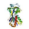
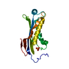
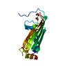
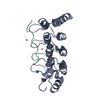
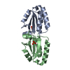
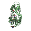
 Deinococcus radiodurans R1 (radioresistant)
 X-RAY DIFFRACTION /
  Authors
 Citation
 Structure visualization
 Downloads & links
 Other downloads


 PDBj
PDBj

 Assembly


 Mass: 22.990 Da / Num. of mol.: 2 / Source method: obtained synthetically / Formula: Na
 Mass: 94.971 Da / Num. of mol.: 2 / Source method: obtained synthetically / Formula: PO4
 Mass: 194.226 Da / Num. of mol.: 1 / Source method: obtained synthetically / Formula: C8H18O5 / Comment: precipitant*YM
 Mass: 92.094 Da / Num. of mol.: 1 / Source method: obtained synthetically / Formula: C3H8O3
 Sample preparation
 / Beamline: BL9-2 / Wavelength: 0.91837, 0.97922, 0.97908
 Processing