 Movie
Movie Controller

[English] 日本語
 Yorodumi
- PDB-3ch9: Crystal structure of Aspergillus fumigatus chitinase B1 in comple... -
+ Open data

- Basic information
| Entry | Database: PDB / ID: 3ch9 | ||||||
|---|---|---|---|---|---|---|---|
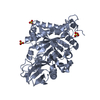
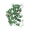
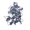
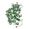
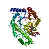
| Title | Crystal structure of Aspergillus fumigatus chitinase B1 in complex with dimethylguanylurea | ||||||
 Components Components | Chitinase | ||||||
 Keywords Keywords | HYDROLASE/HYDROLASE inhibitor / (beta-alpha)8 barrel / chitinase / peptide inhibitors / hydrolase / Glycosidase / HYDROLASE-HYDROLASE inhibitor complex | ||||||
| Function / homology |  Function and homology information Function and homology informationendochitinase activity / chitinase / chitin catabolic process / chitin binding / polysaccharide catabolic process / extracellular region Similarity search - Function | ||||||
| Biological species |  | ||||||
| Method |  X-RAY DIFFRACTION / Known phases / Resolution: 2.2 Å X-RAY DIFFRACTION / Known phases / Resolution: 2.2 Å | ||||||
 Authors Authors | Andersen, O.A. / van Aalten, D.M.F. | ||||||
 Citation Citation | Journal: Chem.Biol. / Year: 2008 Title: Structure-based dissection of the natural product cyclopentapeptide chitinase inhibitor argifin. Authors: Andersen, O.A. / Nathubhai, A. / Dixon, M.J. / Eggleston, I.M. / van Aalten, D.M. | ||||||
| History |
|
- Structure visualization
| Structure viewer | Molecule: MolmilJmol/JSmol |
|---|
- Downloads & links
-Download
| PDBx/mmCIF format | 3ch9.cif.gz | 177.5 KB | Display | PDBx/mmCIF format |
|---|---|---|---|---|
| PDB format | pdb3ch9.ent.gz | 139.9 KB | Display | PDB format |
| PDBx/mmJSON format | 3ch9.json.gz | Tree view | PDBx/mmJSON format | |
| Others |  Other downloads Other downloads |
-Validation report
| Arichive directory | https://data.pdbj.org/pub/pdb/validation_reports/ch/3ch9ftp://data.pdbj.org/pub/pdb/validation_reports/ch/3ch9 | HTTPS FTP |
|---|
-Related structure data
| Related structure data |  3chcC 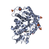 3chdC 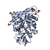 3cheC 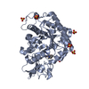 3chfC  1w9vS S: Starting model for refinement C: citing same article ( |
|---|---|
| Similar structure data |


-Links
 PDBj
PDBj- Assembly
| Deposited unit | 
| ||||||||
|---|---|---|---|---|---|---|---|---|---|
| 1 | 
| ||||||||
| 2 | 
| ||||||||
| Unit cell |
|
-Components
| #1: Protein | Mass: 47661.836 Da / Num. of mol.: 2 Source method: isolated from a genetically manipulated source Source: (gene. exp.)  #2: Chemical | ChemComp-SO4 /   Mass: 96.063 Da / Num. of mol.: 13 / Source method: obtained synthetically / Formula: SO4 Mass: 96.063 Da / Num. of mol.: 13 / Source method: obtained synthetically / Formula: SO4#3: Chemical |   Mass: 130.148 Da / Num. of mol.: 2 / Source method: obtained synthetically / Formula: C4H10N4O Mass: 130.148 Da / Num. of mol.: 2 / Source method: obtained synthetically / Formula: C4H10N4O#4: Water | ChemComp-HOH / |  Mass: 18.015 Da / Num. of mol.: 552 / Source method: isolated from a natural source / Formula: H2O Mass: 18.015 Da / Num. of mol.: 552 / Source method: isolated from a natural source / Formula: H2O |
|---|
-Experimental details
-Experiment
| Experiment | Method: X-RAY DIFFRACTION / Number of used crystals: 1 |
|---|
- Sample preparation
| Crystal | Density Matthews: 3.62 Å3/Da / Density % sol: 66.04 % |
|---|---|
| Crystal grow | Temperature: 293 K / Method: vapor diffusion, hanging drop / pH: 9.5 Details: Tris/HCl, Li2SO4, pH 9.5, VAPOR DIFFUSION, HANGING DROP, temperature 293K |
-Data collection
| Diffraction | Mean temperature: 173 K |
|---|---|
| Diffraction source | Source: ROTATING ANODE / Type: RIGAKU / Wavelength: 1.5418 Å |
| Detector | Type: RIGAKU RAXIS IV / Detector: IMAGE PLATE / Date: Aug 30, 2007 / Details: mirrors |
| Radiation | Monochromator: mirrors / Protocol: SINGLE WAVELENGTH / Monochromatic (M) / Laue (L): M / Scattering type: x-ray |
| Radiation wavelength | Wavelength: 1.5418 Å / Relative weight: 1 |
| Reflection | Resolution: 2.2→20 Å / Num. all: 68966 / Num. obs: 67434 / % possible obs: 97.9 % / Observed criterion σ(F): 0 / Observed criterion σ(I): 0 / Redundancy: 3.3 % / Biso Wilson estimate: 37.9 Å2 / Rmerge(I) obs: 0.084 / Net I/σ(I): 17.5 |
| Reflection shell | Resolution: 2.2→2.28 Å / Redundancy: 2.8 % / Rmerge(I) obs: 0.492 / Mean I/σ(I) obs: 2.1 / % possible all: 95.6 |
- Processing
| Software |
| |||||||||||||||||||||||||
|---|---|---|---|---|---|---|---|---|---|---|---|---|---|---|---|---|---|---|---|---|---|---|---|---|---|---|
| Refinement | Method to determine structure: Known phases Starting model: PDB entry 1W9V Resolution: 2.2→20 Å / Isotropic thermal model: Isotropic / Cross valid method: THROUGHOUT / σ(F): 0 / σ(I): 0 / Stereochemistry target values: Engh & Huber
| |||||||||||||||||||||||||
| Displacement parameters | Biso mean: 33.3 Å2 | |||||||||||||||||||||||||
| Refine analyze |
| |||||||||||||||||||||||||
| Refinement step | Cycle: LAST / Resolution: 2.2→20 Å
| |||||||||||||||||||||||||
| Refine LS restraints |
| |||||||||||||||||||||||||
| LS refinement shell | Resolution: 2.2→2.26 Å
|
