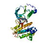
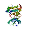
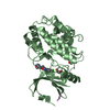
[English] 日本語
Yorodumi
- PDB-3cc6: Crystal structure of kinase domain of protein tyrosine kinase 2 b... -

+
Open data


ID or keywords:

Loading...

-
Basic information

Entry
Database: PDB / ID: 3cc6
TitleCrystal structure of kinase domain of protein tyrosine kinase 2 beta (PTK2B)
ComponentsProtein tyrosine kinase 2 beta
KeywordsTRANSFERASE / FOCAL ADHESION KINASE / Structural Genomics / Structural Genomics Consortium / SGC / ATP-binding / Membrane / Nucleotide-binding / Phosphoprotein / Tyrosine-protein kinase
Function / homology
Function and homology information


regulation of macrophage chemotaxis / neurotransmitter receptor regulator activity / response to cation stress / positive regulation of B cell chemotaxis / marginal zone B cell differentiation / 3-phosphoinositide-dependent protein kinase binding / regulation of ubiquitin-dependent protein catabolic process / endothelin receptor signaling pathway / negative regulation of myeloid cell differentiation / regulation of postsynaptic density assembly ...regulation of macrophage chemotaxis / neurotransmitter receptor regulator activity / response to cation stress / positive regulation of B cell chemotaxis / marginal zone B cell differentiation / 3-phosphoinositide-dependent protein kinase binding / regulation of ubiquitin-dependent protein catabolic process / endothelin receptor signaling pathway / negative regulation of myeloid cell differentiation / regulation of postsynaptic density assembly / blood vessel endothelial cell migration / negative regulation of muscle cell apoptotic process / negative regulation of bone mineralization / cortical cytoskeleton organization / apical dendrite / positive regulation of ubiquitin-dependent protein catabolic process / activation of Janus kinase activity / regulation of release of sequestered calcium ion into cytosol / cellular response to fluid shear stress / focal adhesion assembly / signal complex assembly / chemokine-mediated signaling pathway / NMDA selective glutamate receptor complex / Interleukin-2 signaling / sprouting angiogenesis / long-term synaptic depression / oocyte maturation / Signal regulatory protein family interactions / positive regulation of cell-matrix adhesion / calcium/calmodulin-dependent protein kinase activity / positive regulation of DNA biosynthetic process / postsynaptic modulation of chemical synaptic transmission / positive regulation of actin filament polymerization / stress fiber assembly / negative regulation of potassium ion transport / positive regulation of excitatory postsynaptic potential / RHOU GTPase cycle / response to immobilization stress / positive regulation of protein kinase activity / postsynaptic density, intracellular component / glial cell proliferation / glutamate receptor binding / cellular defense response / regulation of cell adhesion / vascular endothelial growth factor receptor signaling pathway / response to glucose / response to mechanical stimulus / cellular response to retinoic acid / peptidyl-tyrosine autophosphorylation / bone resorption / response to cAMP / tumor necrosis factor-mediated signaling pathway / ionotropic glutamate receptor signaling pathway / positive regulation of endothelial cell migration / positive regulation of synaptic transmission, glutamatergic / response to hormone / response to cocaine / positive regulation of translation / response to ischemia / integrin-mediated signaling pathway / long-term synaptic potentiation / regulation of actin cytoskeleton organization / positive regulation of JNK cascade / non-specific protein-tyrosine kinase / non-membrane spanning protein tyrosine kinase activity / Schaffer collateral - CA1 synapse / regulation of synaptic plasticity / response to hydrogen peroxide / epidermal growth factor receptor signaling pathway / VEGFA-VEGFR2 Pathway / positive regulation of neuron projection development / response to calcium ion / peptidyl-tyrosine phosphorylation / positive regulation of angiogenesis / : / positive regulation of reactive oxygen species metabolic process / neuron projection development / MAPK cascade / positive regulation of nitric oxide biosynthetic process / presynapse / lamellipodium / cell cortex / regulation of cell shape / cell body / positive regulation of cytosolic calcium ion concentration / growth cone / positive regulation of cell growth / protein-containing complex assembly / protein tyrosine kinase activity / response to ethanol / adaptive immune response / negative regulation of neuron apoptotic process / protein autophosphorylation / dendritic spine / postsynaptic density / positive regulation of phosphatidylinositol 3-kinase/protein kinase B signal transduction / cell surface receptor signaling pathway / positive regulation of ERK1 and ERK2 cascade / cytoskeleton / response to hypoxia
Similarity search - Function
Focal adhesion kinase, targeting (FAT) domain / Focal adhesion kinase, targeting (FAT) domain superfamily / Focal adhesion kinase, N-terminal / FAK1/PYK2, FERM domain C-lobe / Focal adhesion targeting region / FERM N-terminal domain / : / FAK1/PYK2, FERM domain C-lobe / FERM central domain / FERM/acyl-CoA-binding protein superfamily ...Focal adhesion kinase, targeting (FAT) domain / Focal adhesion kinase, targeting (FAT) domain superfamily / Focal adhesion kinase, N-terminal / FAK1/PYK2, FERM domain C-lobe / Focal adhesion targeting region / FERM N-terminal domain / : / FAK1/PYK2, FERM domain C-lobe / FERM central domain / FERM/acyl-CoA-binding protein superfamily / FERM central domain / FERM superfamily, second domain / FERM domain / FERM domain profile. / Band 4.1 domain / Band 4.1 homologues / Tyrosine-protein kinase, catalytic domain / Tyrosine kinase, catalytic domain / Tyrosine protein kinases specific active-site signature. / PH-like domain superfamily / Tyrosine-protein kinase, active site / Protein tyrosine and serine/threonine kinase / Serine-threonine/tyrosine-protein kinase, catalytic domain / Ubiquitin-like domain superfamily / Transferase(Phosphotransferase) domain 1 / Transferase(Phosphotransferase); domain 1 / Phosphorylase Kinase; domain 1 / Phosphorylase Kinase; domain 1 / Protein kinase, ATP binding site / Protein kinases ATP-binding region signature. / Protein kinase domain profile. / Protein kinase domain / Protein kinase-like domain superfamily / 2-Layer Sandwich / Orthogonal Bundle / Mainly Alpha / Alpha Beta
Similarity search - Domain/homology
Protein-tyrosine kinase 2-beta
Similarity search - Component
Biological speciesHomo sapiens (human)
MethodX-RAY DIFFRACTION / SYNCHROTRON / MOLECULAR REPLACEMENT / molecular replacement / Resolution: 1.6 Å
AuthorsBusam, R.D. / Lehtio, L. / Karlberg, T. / Arrowsmith, C.H. / Bountra, C. / Collins, R. / Dahlgren, L.G. / Edwards, A.M. / Flodin, S. / Flores, A. ...Busam, R.D. / Lehtio, L. / Karlberg, T. / Arrowsmith, C.H. / Bountra, C. / Collins, R. / Dahlgren, L.G. / Edwards, A.M. / Flodin, S. / Flores, A. / Graslund, S. / Hammarstrom, M. / Helleday, T. / Herman, M.D. / Johansson, A. / Johansson, I. / Kallas, A. / Kotenyova, T. / Moche, M. / Nilsson, M.E. / Nordlund, P. / Nyman, T. / Persson, C. / Sagemark, J. / Svensson, L. / Thorsell, A.G. / Tresaugues, L. / Van den Berg, S. / Weigelt, J. / Welin, M. / Berglund, H. / Structural Genomics Consortium (SGC)
CitationJournal: To be Published
Title: Structure of Protein Tyrosine Kinase 2 Beta (PTK2B) Kinase domain.
Authors: Busam, R.D. / Lehtio, L. / Karlberg, T. / Arrowsmith, C.H. / Bountra, C. / Collins, R. / Dahlgren, L.G. / Edwards, A.M. / Flodin, S. / Flores, A. / Graslund, S. / Hammarstrom, M. / Helleday, ...Authors: Busam, R.D. / Lehtio, L. / Karlberg, T. / Arrowsmith, C.H. / Bountra, C. / Collins, R. / Dahlgren, L.G. / Edwards, A.M. / Flodin, S. / Flores, A. / Graslund, S. / Hammarstrom, M. / Helleday, T. / Herman, M.D. / Johansson, A. / Johansson, I. / Kallas, A. / Kotenyova, T. / Moche, M. / Nilsson, M.E. / Nordlund, P. / Nyman, T. / Persson, C. / Sagemark, J. / Svensson, L. / Thorsell, A.G. / Tresaugues, L. / Van den Berg, S. / Weigelt, J. / Welin, M. / Berglund, H.
History
DepositionFeb 25, 2008Deposition site: RCSB / Processing site: RCSB
Revision 1.0Mar 11, 2008Provider: repository / Type: Initial release
Revision 1.1Jul 13, 2011Group: Non-polymer description / Version format compliance
Revision 1.2Aug 30, 2023Group: Data collection / Database references ...Data collection / Database references / Derived calculations / Refinement description
Category: chem_comp_atom / chem_comp_bond ...chem_comp_atom / chem_comp_bond / database_2 / pdbx_initial_refinement_model / pdbx_struct_conn_angle / struct_conn / struct_ref_seq_dif / struct_site
Item: _database_2.pdbx_DOI / _database_2.pdbx_database_accession ..._database_2.pdbx_DOI / _database_2.pdbx_database_accession / _pdbx_struct_conn_angle.ptnr1_auth_comp_id / _pdbx_struct_conn_angle.ptnr1_auth_seq_id / _pdbx_struct_conn_angle.ptnr1_label_asym_id / _pdbx_struct_conn_angle.ptnr1_label_atom_id / _pdbx_struct_conn_angle.ptnr1_label_comp_id / _pdbx_struct_conn_angle.ptnr1_label_seq_id / _pdbx_struct_conn_angle.ptnr3_auth_comp_id / _pdbx_struct_conn_angle.ptnr3_auth_seq_id / _pdbx_struct_conn_angle.ptnr3_label_asym_id / _pdbx_struct_conn_angle.ptnr3_label_atom_id / _pdbx_struct_conn_angle.ptnr3_label_comp_id / _pdbx_struct_conn_angle.ptnr3_label_seq_id / _pdbx_struct_conn_angle.value / _struct_conn.pdbx_dist_value / _struct_conn.ptnr1_auth_comp_id / _struct_conn.ptnr1_auth_seq_id / _struct_conn.ptnr1_label_asym_id / _struct_conn.ptnr1_label_atom_id / _struct_conn.ptnr1_label_comp_id / _struct_conn.ptnr1_label_seq_id / _struct_conn.ptnr2_auth_comp_id / _struct_conn.ptnr2_auth_seq_id / _struct_conn.ptnr2_label_asym_id / _struct_conn.ptnr2_label_atom_id / _struct_conn.ptnr2_label_comp_id / _struct_ref_seq_dif.details / _struct_site.pdbx_auth_asym_id / _struct_site.pdbx_auth_comp_id / _struct_site.pdbx_auth_seq_id

-
Structure visualization

Structure viewerMolecule:
MolmilJmol/JSmol

Downloads & links

-
Assembly

Deposited unit
A: Protein tyrosine kinase 2 beta
hetero molecules


Theoretical massNumber of molelcules
Total (without water)32,6393
Polymers32,5231
Non-polymers1162
Water4,035224
1


  • Idetical with deposited unit
  • defined by author&software
TypeNameSymmetry operationNumber
identity operation1_555x,y,z1
MethodPISA
Unit cell
Length a, b, c (Å)37.450, 96.140, 43.200
Angle α, β, γ (deg.)90.000, 93.660, 90.000
Int Tables number4
Space group name H-MP1211

-
Components

#1: Protein Protein tyrosine kinase 2 beta / Focal adhesion kinase 2 / FADK 2 / Proline-rich tyrosine kinase 2 / Cell adhesion kinase beta / CAK ...Focal adhesion kinase 2 / FADK 2 / Proline-rich tyrosine kinase 2 / Cell adhesion kinase beta / CAK beta / Calcium-dependent tyrosine kinase / CADTK / Related adhesion focal tyrosine kinase / RAFTK


Mass: 32522.701 Da / Num. of mol.: 1 / Fragment: Kinase domain: Residues 414-692
Source method: isolated from a genetically manipulated source
Source: (gene. exp.) Homo sapiens (human) / Gene: PTK2B, FAK2, PYK2, RAFTK / Plasmid: pNIC-Bsa4 / Production host: Escherichia coli (E. coli) / Strain (production host): BL21(DE3) gold YopH
References: UniProt: Q14289, non-specific protein-tyrosine kinase
#2: Chemical ChemComp-MG / MAGNESIUM ION


Mass: 24.305 Da / Num. of mol.: 1 / Source method: obtained synthetically / Formula: Mg
#3: Chemical ChemComp-GOL / GLYCEROL / GLYCERIN / PROPANE-1,2,3-TRIOL


Mass: 92.094 Da / Num. of mol.: 1 / Source method: obtained synthetically / Formula: C3H8O3
#4: Water ChemComp-HOH / water


Mass: 18.015 Da / Num. of mol.: 224 / Source method: isolated from a natural source / Formula: H2O

-
Experimental details

-
Experiment

ExperimentMethod: X-RAY DIFFRACTION / Number of used crystals: 1

-
Sample preparation

CrystalDensity Matthews: 2.39 Å3/Da / Density % sol: 48.46 %
Crystal growTemperature: 277 K / Method: vapor diffusion, sitting drop / pH: 5.5
Details: 0.05M Magnesium chloride, 0.1M Bis-tris, 17.5% PEG 3350, pH 5.5, VAPOR DIFFUSION, SITTING DROP, temperature 277K

-
Data collection

DiffractionMean temperature: 100 K
Diffraction sourceSource: SYNCHROTRON / Site: ESRF / Beamline: ID29 / Wavelength: 0.9753 Å
DetectorType: ADSC QUANTUM 315 / Detector: CCD / Date: Sep 13, 2007
RadiationMonochromator: Si(111) / Protocol: SINGLE WAVELENGTH / Monochromatic (M) / Laue (L): M / Scattering type: x-ray
Radiation wavelengthWavelength: 0.9753 Å / Relative weight: 1
ReflectionResolution: 1.6→18.84 Å / Num. all: 49357 / Num. obs: 40123 / % possible obs: 99.9 % / Observed criterion σ(I): -3 / Redundancy: 7.4 % / Biso Wilson estimate: 22.575 Å2 / Rmerge(I) obs: 0.081 / Net I/σ(I): 16.22
Reflection shellResolution: 1.6→1.7 Å / Rmerge(I) obs: 0.441 / Mean I/σ(I) obs: 4.6 / Num. unique obs: 6650 / % possible all: 100

-
Phasing

PhasingMethod: molecular replacement

-
Processing

Software
NameVersionClassificationNB
XSCALEdata scaling
MOLREPphasing
REFMACrefinement
PDB_EXTRACT3.004data extraction
MxCuBEdata collection
XDSdata reduction
RefinementMethod to determine structure: MOLECULAR REPLACEMENT
Starting model: PDB entry 2ETM
Resolution: 1.6→18.84 Å / Cor.coef. Fo:Fc: 0.953 / Cor.coef. Fo:Fc free: 0.933 / SU B: 1.597 / SU ML: 0.058 / Cross valid method: THROUGHOUT / σ(F): 0 / ESU R: 0.093 / ESU R Free: 0.095 / Stereochemistry target values: MAXIMUM LIKELIHOOD / Details: HYDROGENS HAVE BEEN ADDED IN THE RIDING POSITIONS
RfactorNum. reflection% reflectionSelection details
Rfree0.219 3209 8 %RANDOM
Rwork0.184 ---
all0.187 40120 --
obs0.187 40120 100 %-
Solvent computationIon probe radii: 0.8 Å / Shrinkage radii: 0.8 Å / VDW probe radii: 1.2 Å / Solvent model: MASK
Displacement parametersBiso mean: 20.195 Å2
Baniso -1Baniso -2Baniso -3
1--0.01 Å20 Å20.01 Å2
2--0.01 Å20 Å2
3---0 Å2
Refinement stepCycle: LAST / Resolution: 1.6→18.84 Å
ProteinNucleic acidLigandSolventTotal
Num. atoms2183 0 7 224 2414
Refine LS restraints
Refine-IDTypeDev idealDev ideal targetNumber
X-RAY DIFFRACTIONr_bond_refined_d0.010.0222383
X-RAY DIFFRACTIONr_angle_refined_deg1.2791.9733251
X-RAY DIFFRACTIONr_dihedral_angle_1_deg5.0865301
X-RAY DIFFRACTIONr_dihedral_angle_2_deg33.94323.922102
X-RAY DIFFRACTIONr_dihedral_angle_3_deg14.47215435
X-RAY DIFFRACTIONr_dihedral_angle_4_deg18.0011514
X-RAY DIFFRACTIONr_chiral_restr0.0860.2356
X-RAY DIFFRACTIONr_gen_planes_refined0.0050.021817
X-RAY DIFFRACTIONr_nbd_refined0.2110.21155
X-RAY DIFFRACTIONr_nbtor_refined0.3070.21687
X-RAY DIFFRACTIONr_xyhbond_nbd_refined0.1250.2199
X-RAY DIFFRACTIONr_metal_ion_refined0.0410.22
X-RAY DIFFRACTIONr_symmetry_vdw_refined0.1510.243
X-RAY DIFFRACTIONr_symmetry_hbond_refined0.2330.215
X-RAY DIFFRACTIONr_mcbond_it1.23321487
X-RAY DIFFRACTIONr_mcangle_it1.98432378
X-RAY DIFFRACTIONr_scbond_it1.30521029
X-RAY DIFFRACTIONr_scangle_it1.9953873
LS refinement shellResolution: 1.6→1.641 Å / Total num. of bins used: 20
RfactorNum. reflection% reflection
Rfree0.26 235 -
Rwork0.214 2705 -
all-2940 -
obs--100 %

+
About Yorodumi

-
News

-
Feb 9, 2022. New format data for meta-information of EMDB entries

New format data for meta-information of EMDB entries

  • Version 3 of the EMDB header file is now the official format.
  • The previous official version 1.9 will be removed from the archive.

Related info.:EMDB header

External links:wwPDB to switch to version 3 of the EMDB data model

-
Aug 12, 2020. Covid-19 info

Covid-19 info

URL: https://pdbjlvh1.pdbj.org/emnavi/covid19.php

New page: Covid-19 featured information page in EM Navigator.

Related info.:Covid-19 info / Mar 5, 2020. Novel coronavirus structure data

+
Mar 5, 2020. Novel coronavirus structure data

Novel coronavirus structure data

Related info.:Yorodumi Speices / Aug 12, 2020. Covid-19 info

External links:COVID-19 featured content - PDBj / Molecule of the Month (242):Coronavirus Proteases

+
Jan 31, 2019. EMDB accession codes are about to change! (news from PDBe EMDB page)

EMDB accession codes are about to change! (news from PDBe EMDB page)

  • The allocation of 4 digits for EMDB accession codes will soon come to an end. Whilst these codes will remain in use, new EMDB accession codes will include an additional digit and will expand incrementally as the available range of codes is exhausted. The current 4-digit format prefixed with “EMD-” (i.e. EMD-XXXX) will advance to a 5-digit format (i.e. EMD-XXXXX), and so on. It is currently estimated that the 4-digit codes will be depleted around Spring 2019, at which point the 5-digit format will come into force.
  • The EM Navigator/Yorodumi systems omit the EMD- prefix.

Related info.:Q: What is EMD? / ID/Accession-code notation in Yorodumi/EM Navigator

External links:EMDB Accession Codes are Changing Soon! / Contact to PDBj

+
Jul 12, 2017. Major update of PDB

Major update of PDB

  • wwPDB released updated PDB data conforming to the new PDBx/mmCIF dictionary.
  • This is a major update changing the version number from 4 to 5, and with Remediation, in which all the entries are updated.
  • In this update, many items about electron microscopy experimental information are reorganized (e.g. em_software).
  • Now, EM Navigator and Yorodumi are based on the updated data.

External links:wwPDB Remediation / Enriched Model Files Conforming to OneDep Data Standards Now Available in the PDB FTP Archive

-
Yorodumi

Thousand views of thousand structures

  • Yorodumi is a browser for structure data from EMDB, PDB, SASBDB, etc.
  • This page is also the successor to EM Navigator detail page, and also detail information page/front-end page for Omokage search.
  • The word "yorodu" (or yorozu) is an old Japanese word meaning "ten thousand". "mi" (miru) is to see.

Related info.:EMDB / PDB / SASBDB / Comparison of 3 databanks / Yorodumi Search / Aug 31, 2016. New EM Navigator & Yorodumi / Yorodumi Papers / Jmol/JSmol / Function and homology information / Changes in new EM Navigator and Yorodumi

Read more