 Movie
Movie Controller

[English] 日本語
 Yorodumi
- PDB-3cb7: The crystallographic structure of the digestive lysozyme 2 from M... -
+ Open data

- Basic information
| Entry | Database: PDB / ID: 3cb7 | ||||||
|---|---|---|---|---|---|---|---|
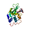
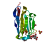
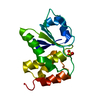
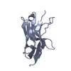
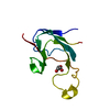
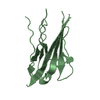
| Title | The crystallographic structure of the digestive lysozyme 2 from Musca domestica at 1.9 Ang. | ||||||
 Components Components | Lys-rich lysozyme 2 | ||||||
 Keywords Keywords | HYDROLASE / Digestive lysozyme 2 / Musca domestica / Glycosidase | ||||||
| Function / homology |  Function and homology information Function and homology information | ||||||
| Biological species |  Musca domestica (house fly) Musca domestica (house fly) | ||||||
| Method |  X-RAY DIFFRACTION / SYNCHROTRON / MOLECULAR REPLACEMENT / Resolution: 1.9 Å X-RAY DIFFRACTION / SYNCHROTRON / MOLECULAR REPLACEMENT / Resolution: 1.9 Å | ||||||
 Authors Authors | Cancado, F.C. / Valerio, A.A. / Marana, S.R. / Barbosa, J.A.R.G. | ||||||
 Citation Citation | Journal: To be Published Title: The crystallographic structure of the digestive lysozyme 2 from Musca domestica at 1.9 Ang. Authors: Marana, S.R. / Cancado, F.C. / Valerio, A.A. / Ferreira, C. / Terra, W.R. / Barbosa, J.A.R.G. #1: Journal: Acta Crystallogr.,Sect.F / Year: 2006Title: Crystallization, data collection and phasing of two digestive lysozymes from Musca domestica Authors: Marana, S.R. / Cancado, F.C. / Valerio, A.A. / Ferreira, C. / Terra, W.R. / Barbosa, J.A.R.G. | ||||||
| History |
|
- Structure visualization
| Structure viewer | Molecule: MolmilJmol/JSmol |
|---|
- Downloads & links
-Download
| PDBx/mmCIF format | 3cb7.cif.gz | 68.9 KB | Display | PDBx/mmCIF format |
|---|---|---|---|---|
| PDB format | pdb3cb7.ent.gz | 51.1 KB | Display | PDB format |
| PDBx/mmJSON format | 3cb7.json.gz | Tree view | PDBx/mmJSON format | |
| Others |  Other downloads Other downloads |
-Validation report
| Arichive directory | https://data.pdbj.org/pub/pdb/validation_reports/cb/3cb7ftp://data.pdbj.org/pub/pdb/validation_reports/cb/3cb7 | HTTPS FTP |
|---|
-Related structure data
| Related structure data |  2fbdS S: Starting model for refinement |
|---|---|
| Similar structure data |


-Links
 PDBj
PDBj


- Assembly
| Deposited unit | 
| ||||||||
|---|---|---|---|---|---|---|---|---|---|
| 1 | 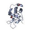
| ||||||||
| 2 | 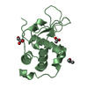
| ||||||||
| Unit cell |
|
-Components
| #1: Protein | Mass: 14305.049 Da / Num. of mol.: 2 Source method: isolated from a genetically manipulated source Source: (gene. exp.) Musca domestica (house fly) / Plasmid: pPIC9 / Production host:  Pichia pastoris (fungus) / Strain (production host): GS115 / References: UniProt: Q7YT17, lysozyme Pichia pastoris (fungus) / Strain (production host): GS115 / References: UniProt: Q7YT17, lysozyme#2: Chemical |   Mass: 60.095 Da / Num. of mol.: 2 / Source method: obtained synthetically / Formula: C3H8O Mass: 60.095 Da / Num. of mol.: 2 / Source method: obtained synthetically / Formula: C3H8O#3: Chemical |   Mass: 60.052 Da / Num. of mol.: 3 / Source method: obtained synthetically / Formula: C2H4O2 Mass: 60.052 Da / Num. of mol.: 3 / Source method: obtained synthetically / Formula: C2H4O2#4: Chemical | ChemComp-GOL / |   Mass: 92.094 Da / Num. of mol.: 1 / Source method: obtained synthetically / Formula: C3H8O3 Mass: 92.094 Da / Num. of mol.: 1 / Source method: obtained synthetically / Formula: C3H8O3#5: Water | ChemComp-HOH / |  Mass: 18.015 Da / Num. of mol.: 279 / Source method: isolated from a natural source / Formula: H2O Mass: 18.015 Da / Num. of mol.: 279 / Source method: isolated from a natural source / Formula: H2OHas protein modification | Y | |
|---|
-Experimental details
-Experiment
| Experiment | Method: X-RAY DIFFRACTION / Number of used crystals: 1 |
|---|
- Sample preparation
| Crystal | Density Matthews: 2.07 Å3/Da / Density % sol: 40.61 % |
|---|---|
| Crystal grow | Temperature: 291 K / Method: vapor diffusion, sitting drop / pH: 4.2 Details: The 3 uL drop was formed by 1.5 uL of protein solution at 9.8 mg/mL in 10 mM MES pH 5.5 plus 1.3 uL of 28% 2-propanol, 0.115 M sodium citrate pH 4.2 and 21% PEG 4000 plus 0.2 uL of 0.2 M ...Details: The 3 uL drop was formed by 1.5 uL of protein solution at 9.8 mg/mL in 10 mM MES pH 5.5 plus 1.3 uL of 28% 2-propanol, 0.115 M sodium citrate pH 4.2 and 21% PEG 4000 plus 0.2 uL of 0.2 M ammonium acetate, 0.1 M sodium citrate pH 5.6 and 30% MPD., VAPOR DIFFUSION, SITTING DROP, temperature 291K |
-Data collection
| Diffraction | Mean temperature: 100 K |
|---|---|
| Diffraction source | Source: SYNCHROTRON / Site: LNLS  / Beamline: D03B-MX1 / Wavelength: 1.427 Å / Beamline: D03B-MX1 / Wavelength: 1.427 Å |
| Detector | Type: MAR CCD 165 mm / Detector: CCD / Date: Mar 20, 2006 / Details: mirrors |
| Radiation | Monochromator: Si crystal / Protocol: SINGLE WAVELENGTH / Monochromatic (M) / Laue (L): M / Scattering type: x-ray |
| Radiation wavelength | Wavelength: 1.427 Å / Relative weight: 1 |
| Reflection | Resolution: 1.9→30 Å / Num. all: 17698 / Num. obs: 17698 / % possible obs: 90.7 % / Observed criterion σ(F): 0 / Observed criterion σ(I): 0 / Redundancy: 3.9 % / Biso Wilson estimate: 23.3 Å2 / Rmerge(I) obs: 0.089 / Net I/σ(I): 14.4 |
| Reflection shell | Resolution: 1.9→1.97 Å / Redundancy: 1.8 % / Rmerge(I) obs: 0.304 / Mean I/σ(I) obs: 2.1 / Num. unique all: 1423 / % possible all: 74.1 |
- Processing
| Software |
| ||||||||||||||||||||||||||||||||||||||||||||||||||||||||||||||||||||||||||||||||||||||||||||||||||||||||||||||||||||||||||||||||||||||||||||||||||||||||||||||||||||||||||
|---|---|---|---|---|---|---|---|---|---|---|---|---|---|---|---|---|---|---|---|---|---|---|---|---|---|---|---|---|---|---|---|---|---|---|---|---|---|---|---|---|---|---|---|---|---|---|---|---|---|---|---|---|---|---|---|---|---|---|---|---|---|---|---|---|---|---|---|---|---|---|---|---|---|---|---|---|---|---|---|---|---|---|---|---|---|---|---|---|---|---|---|---|---|---|---|---|---|---|---|---|---|---|---|---|---|---|---|---|---|---|---|---|---|---|---|---|---|---|---|---|---|---|---|---|---|---|---|---|---|---|---|---|---|---|---|---|---|---|---|---|---|---|---|---|---|---|---|---|---|---|---|---|---|---|---|---|---|---|---|---|---|---|---|---|---|---|---|---|---|---|---|
| Refinement | Method to determine structure: MOLECULAR REPLACEMENT Starting model: pdb entry 2fbd Resolution: 1.9→30 Å / Cor.coef. Fo:Fc: 0.965 / Cor.coef. Fo:Fc free: 0.923 / SU B: 15.216 / SU ML: 0.209 / TLS residual ADP flag: LIKELY RESIDUAL / Cross valid method: THROUGHOUT / σ(F): 0 / ESU R: 0.203 / ESU R Free: 0.191 / Stereochemistry target values: MAXIMUM LIKELIHOOD
| ||||||||||||||||||||||||||||||||||||||||||||||||||||||||||||||||||||||||||||||||||||||||||||||||||||||||||||||||||||||||||||||||||||||||||||||||||||||||||||||||||||||||||
| Solvent computation | Ion probe radii: 0.8 Å / Shrinkage radii: 0.8 Å / VDW probe radii: 1.2 Å / Solvent model: BABINET MODEL WITH MASK | ||||||||||||||||||||||||||||||||||||||||||||||||||||||||||||||||||||||||||||||||||||||||||||||||||||||||||||||||||||||||||||||||||||||||||||||||||||||||||||||||||||||||||
| Displacement parameters | Biso mean: 40.144 Å2
| ||||||||||||||||||||||||||||||||||||||||||||||||||||||||||||||||||||||||||||||||||||||||||||||||||||||||||||||||||||||||||||||||||||||||||||||||||||||||||||||||||||||||||
| Refinement step | Cycle: LAST / Resolution: 1.9→30 Å
| ||||||||||||||||||||||||||||||||||||||||||||||||||||||||||||||||||||||||||||||||||||||||||||||||||||||||||||||||||||||||||||||||||||||||||||||||||||||||||||||||||||||||||
| Refine LS restraints |
| ||||||||||||||||||||||||||||||||||||||||||||||||||||||||||||||||||||||||||||||||||||||||||||||||||||||||||||||||||||||||||||||||||||||||||||||||||||||||||||||||||||||||||
| LS refinement shell | Resolution: 1.9→1.95 Å / Total num. of bins used: 20
| ||||||||||||||||||||||||||||||||||||||||||||||||||||||||||||||||||||||||||||||||||||||||||||||||||||||||||||||||||||||||||||||||||||||||||||||||||||||||||||||||||||||||||
| Refinement TLS params. | Method: refined / Refine-ID: X-RAY DIFFRACTION
| ||||||||||||||||||||||||||||||||||||||||||||||||||||||||||||||||||||||||||||||||||||||||||||||||||||||||||||||||||||||||||||||||||||||||||||||||||||||||||||||||||||||||||
| Refinement TLS group |
|
