 Movie
Movie Controller

[English] 日本語
 Yorodumi
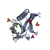
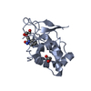
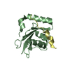
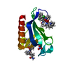
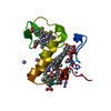
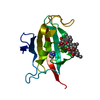
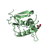
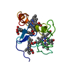
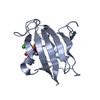
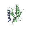
- PDB-3car: REDUCED STRUCTURE OF THE ACIDIC CYTOCHROME C3 FROM DESULFOVIBRIO ... -
+ Open data

- Basic information
| Entry | Database: PDB / ID: 3car | ||||||
|---|---|---|---|---|---|---|---|
| Title | REDUCED STRUCTURE OF THE ACIDIC CYTOCHROME C3 FROM DESULFOVIBRIO AFRICANUS | ||||||
 Components Components | CYTOCHROME C3 | ||||||
 Keywords Keywords | ELECTRON TRANSPORT / CYTOCHROME C3 / TETRAHEME / REDUCED FORM / DESULFOVIBRIO AFRICANUS | ||||||
| Function / homology |  Function and homology information Function and homology informationanaerobic respiration / periplasmic space / electron transfer activity / heme binding / metal ion binding Similarity search - Function | ||||||
| Biological species |  Desulfovibrio africanus (bacteria) Desulfovibrio africanus (bacteria) | ||||||
| Method |  X-RAY DIFFRACTION / SYNCHROTRON / MOLECULAR REPLACEMENT / Resolution: 1.9 Å X-RAY DIFFRACTION / SYNCHROTRON / MOLECULAR REPLACEMENT / Resolution: 1.9 Å | ||||||
 Authors Authors | Norager, S. / Legrand, P. / Pieulle, L. / Hatchikian, C. / Roth, M. | ||||||
 Citation Citation | Journal: J.Mol.Biol. / Year: 1999 Title: Crystal structure of the oxidised and reduced acidic cytochrome c3from Desulfovibrio africanus. Authors: Norager, S. / Legrand, P. / Pieulle, L. / Hatchikian, C. / Roth, M. #1: Journal: Thesis, Universite Joseph Fourier / Year: 1998Title: Crystallographic Studies of the Two Tetraheme Cytochromes C3 from Desulfovibrio Africanus Authors: Norager, S.C. #2: Journal: Biochim.Biophys.Acta / Year: 1997Title: Further Characterization of the Two Tetraheme Cytochromes C3 from Desulfovibrio Africanus: Nucleotide Sequences, Epr Spectroscopy and Biological Activity Authors: Magro, V. / Pieulle, L. / Forget, N. / Guigliarelli, B. / Petillot, Y. / Hatchikian, E.C. #3: Journal: Biochim.Biophys.Acta / Year: 1996Title: Biochemical Studies of the C-Type Cytochromes of the Sulfate Reducer Desulfovibrio Africanus. Characterization of Two Tetraheme Cytochromes C3 with Different Specificity Authors: Pieulle, L. / Haladjian, J. / Bonicel, J. / Hatchikian, E.C. | ||||||
| History |
|
- Structure visualization
| Structure viewer | Molecule: MolmilJmol/JSmol |
|---|
- Downloads & links
-Download
| PDBx/mmCIF format | 3car.cif.gz | 44.9 KB | Display | PDBx/mmCIF format |
|---|---|---|---|---|
| PDB format | pdb3car.ent.gz | 31.5 KB | Display | PDB format |
| PDBx/mmJSON format | 3car.json.gz | Tree view | PDBx/mmJSON format | |
| Others |  Other downloads Other downloads |
-Validation report
| Arichive directory | https://data.pdbj.org/pub/pdb/validation_reports/ca/3carftp://data.pdbj.org/pub/pdb/validation_reports/ca/3car | HTTPS FTP |
|---|
-Related structure data

-Links
 PDBj
PDBj
- Assembly
| Deposited unit | 
| ||||||||
|---|---|---|---|---|---|---|---|---|---|
| 1 |
| ||||||||
| 2 | 
| ||||||||
| Unit cell |
| ||||||||
| Components on special symmetry positions |
|
-Components
| #1: Protein | Mass: 11278.314 Da / Num. of mol.: 1 / Source method: isolated from a natural source / Source: (natural) Desulfovibrio africanus (bacteria) / Cellular location: PERIPLASM / Strain: BENGHAZI / References: UniProt: P94690 | ||||||
|---|---|---|---|---|---|---|---|
| #2: Chemical | ChemComp-ZN /   Mass: 65.409 Da / Num. of mol.: 4 / Source method: obtained synthetically / Formula: Zn Mass: 65.409 Da / Num. of mol.: 4 / Source method: obtained synthetically / Formula: Zn#3: Chemical | ChemComp-HEM /   Mass: 616.487 Da / Num. of mol.: 4 / Source method: obtained synthetically / Formula: C34H32FeN4O4 Mass: 616.487 Da / Num. of mol.: 4 / Source method: obtained synthetically / Formula: C34H32FeN4O4#4: Chemical |   Mass: 74.922 Da / Num. of mol.: 3 / Source method: obtained synthetically / Formula: As Mass: 74.922 Da / Num. of mol.: 3 / Source method: obtained synthetically / Formula: As#5: Water | ChemComp-HOH / |  Mass: 18.015 Da / Num. of mol.: 159 / Source method: isolated from a natural source / Formula: H2O Mass: 18.015 Da / Num. of mol.: 159 / Source method: isolated from a natural source / Formula: H2O |
-Experimental details
-Experiment
| Experiment | Method: X-RAY DIFFRACTION / Number of used crystals: 1 |
|---|
- Sample preparation
| Crystal | Density Matthews: 3.34 Å3/Da / Density % sol: 67 % | |||||||||||||||||||||||||
|---|---|---|---|---|---|---|---|---|---|---|---|---|---|---|---|---|---|---|---|---|---|---|---|---|---|---|
| Crystal grow | pH: 5.8 / Details: pH 5.80 | |||||||||||||||||||||||||
| Crystal grow | *PLUS Method: vapor diffusion, hanging dropDetails: drop consists of equal volume of protein and reservoir solutions PH range low: 5.8 / PH range high: 5.6 | |||||||||||||||||||||||||
| Components of the solutions | *PLUS
|
-Data collection
| Diffraction | Mean temperature: 108.2 K |
|---|---|
| Diffraction source | Source: SYNCHROTRON / Site: ESRF  / Beamline: BM02 / Wavelength: 1.0057 / Beamline: BM02 / Wavelength: 1.0057 |
| Detector | Type: THOMSON/PRINCETON JNSJR / Detector: CCD AREA DETECTOR / Date: Jan 21, 1998 |
| Radiation | Protocol: SINGLE WAVELENGTH / Monochromatic (M) / Laue (L): M / Scattering type: x-ray |
| Radiation wavelength | Wavelength: 1.0057 Å / Relative weight: 1 |
| Reflection | Resolution: 1.85→29 Å / Num. obs: 17501 / % possible obs: 97.9 % / Redundancy: 32.4 % / Biso Wilson estimate: 21.12 Å2 / Rsym value: 0.065 / Net I/σ(I): 8.6 |
| Reflection shell | Resolution: 1.85→1.95 Å / Redundancy: 13.8 % / Mean I/σ(I) obs: 1.2 / Rsym value: 0.531 / % possible all: 87.4 |
| Reflection | *PLUS Rmerge(I) obs: 0.065 |
| Reflection shell | *PLUS % possible obs: 87.4 % / Rmerge(I) obs: 0.531 |
- Processing
| Software |
| |||||||||||||||||||||||||||||||||||||||||||||||||||||||||||||||
|---|---|---|---|---|---|---|---|---|---|---|---|---|---|---|---|---|---|---|---|---|---|---|---|---|---|---|---|---|---|---|---|---|---|---|---|---|---|---|---|---|---|---|---|---|---|---|---|---|---|---|---|---|---|---|---|---|---|---|---|---|---|---|---|---|
| Refinement | Method to determine structure: MOLECULAR REPLACEMENT Starting model: OXIDISED FORM OF THE SAME PROTEIN Resolution: 1.9→20 Å / Cross valid method: THROUGHOUT / σ(F): 0 / ESU R: 0.15 Details: RESIDUES WITH AN OCCUPANCY LOWER THAN 1.0, ARE REFINED WITH THE GIVEN OCCUPANCY. THIS OCCUPANCY HAS BEEN ESTABLISHED MANUALLY TO AVOID POSITIVE OR NEGATIVE PEAKS IN THE FOBS-FCALC DENSITY MAPS.
| |||||||||||||||||||||||||||||||||||||||||||||||||||||||||||||||
| Displacement parameters | Biso mean: 29.75 Å2 | |||||||||||||||||||||||||||||||||||||||||||||||||||||||||||||||
| Refinement step | Cycle: LAST / Resolution: 1.9→20 Å
| |||||||||||||||||||||||||||||||||||||||||||||||||||||||||||||||
| Refine LS restraints |
|
