 Movie
Movie Controller

[English] 日本語
 Yorodumi
- PDB-3c90: The 1.25 A Resolution Structure of Phosphoribosyl-ATP Pyrophospho... -
+ Open data

- Basic information
| Entry | Database: PDB / ID: 3c90 | ||||||
|---|---|---|---|---|---|---|---|
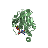
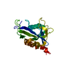
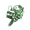
| Title | The 1.25 A Resolution Structure of Phosphoribosyl-ATP Pyrophosphohydrolase from Mycobacterium tuberculosis, crystal form II | ||||||
 Components Components | Phosphoribosyl-ATP pyrophosphatase | ||||||
 Keywords Keywords | HYDROLASE / alpha-helical / Amino-acid biosynthesis / Histidine biosynthesis / Structural Genomics / TB Structural Genomics Consortium / TBSGC / PSI-2 / Protein Structure Initiative | ||||||
| Function / homology |  Function and homology information Function and homology informationresponse to host iron concentration / phosphoribosyl-ATP diphosphatase / phosphoribosyl-ATP diphosphatase activity / cell wall / L-histidine biosynthetic process / peptidoglycan-based cell wall / magnesium ion binding / ATP binding / cytoplasm Similarity search - Function | ||||||
| Biological species |   Mycobacterium tuberculosis (bacteria) Mycobacterium tuberculosis (bacteria) | ||||||
| Method |  X-RAY DIFFRACTION / MOLECULAR REPLACEMENT / Resolution: 1.79 Å X-RAY DIFFRACTION / MOLECULAR REPLACEMENT / Resolution: 1.79 Å | ||||||
 Authors Authors | Javid-Majd, F. / Yang, D. / Ioerger, T.R. / Sacchettini, J.C. / TB Structural Genomics Consortium (TBSGC) | ||||||
 Citation Citation | Journal: Acta Crystallogr.,Sect.D / Year: 2008 Title: The 1.25 A resolution structure of phosphoribosyl-ATP pyrophosphohydrolase from Mycobacterium tuberculosis. Authors: Javid-Majd, F. / Yang, D. / Ioerger, T.R. / Sacchettini, J.C. | ||||||
| History |
|
- Structure visualization
| Structure viewer | Molecule: MolmilJmol/JSmol |
|---|
- Downloads & links
-Download
| PDBx/mmCIF format | 3c90.cif.gz | 81.6 KB | Display | PDBx/mmCIF format |
|---|---|---|---|---|
| PDB format | pdb3c90.ent.gz | 62.8 KB | Display | PDB format |
| PDBx/mmJSON format | 3c90.json.gz | Tree view | PDBx/mmJSON format | |
| Others |  Other downloads Other downloads |
-Validation report
| Arichive directory | https://data.pdbj.org/pub/pdb/validation_reports/c9/3c90ftp://data.pdbj.org/pub/pdb/validation_reports/c9/3c90 | HTTPS FTP |
|---|
-Related structure data
| Related structure data |  1y6xSC S: Starting model for refinement C: citing same article ( |
|---|---|
| Similar structure data | |
| Other databases |


-Links
 PDBj
PDBj- Assembly
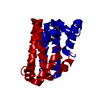
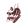
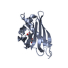
| Deposited unit | 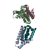
| ||||||||
|---|---|---|---|---|---|---|---|---|---|
| 1 | 
| ||||||||
| 2 | 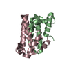
| ||||||||
| Unit cell |
|
-Components
| #1: Protein | Mass: 10154.372 Da / Num. of mol.: 4 Source method: isolated from a genetically manipulated source Source: (gene. exp.) Mycobacterium tuberculosis (bacteria) / Strain: H37Rv / Gene: hisE, Rv2122c, MT2182, MTCY261.18 / Plasmid: pET23a(+) / Production host: References: UniProt: P0A5B1, UniProt: P9WMM9*PLUS, phosphoribosyl-ATP diphosphatase #2: Water | ChemComp-HOH / |  Mass: 18.015 Da / Num. of mol.: 233 / Source method: isolated from a natural source / Formula: H2O Mass: 18.015 Da / Num. of mol.: 233 / Source method: isolated from a natural source / Formula: H2O |
|---|
-Experimental details
-Experiment
| Experiment | Method: X-RAY DIFFRACTION / Number of used crystals: 1 |
|---|
- Sample preparation
| Crystal | Density Matthews: 2.1 Å3/Da / Density % sol: 41.34 % |
|---|---|
| Crystal grow | Temperature: 293 K / Method: vapor diffusion, hanging drop Details: 1M Sodium formate, 0.1M Sodium iodide, 0.1 mM Calcium chloride, VAPOR DIFFUSION, HANGING DROP, temperature 293K |
-Data collection
| Diffraction | Mean temperature: 100 K |
|---|---|
| Diffraction source | Source: ROTATING ANODE / Type: MACSCIENCE / Wavelength: 1.54178 Å |
| Detector | Type: MAC Science DIP-2030 / Detector: IMAGE PLATE / Details: Osmic optics |
| Radiation | Protocol: SINGLE WAVELENGTH / Monochromatic (M) / Laue (L): M / Scattering type: x-ray |
| Radiation wavelength | Wavelength: 1.54178 Å / Relative weight: 1 |
| Reflection | Resolution: 1.79→50 Å / Num. obs: 31912 / % possible obs: 98.9 % |
- Processing
| Software |
| ||||||||||||||||||||||||||||||||||||||||||||||||||||||||||||||||||||||||||||||||||||||||||
|---|---|---|---|---|---|---|---|---|---|---|---|---|---|---|---|---|---|---|---|---|---|---|---|---|---|---|---|---|---|---|---|---|---|---|---|---|---|---|---|---|---|---|---|---|---|---|---|---|---|---|---|---|---|---|---|---|---|---|---|---|---|---|---|---|---|---|---|---|---|---|---|---|---|---|---|---|---|---|---|---|---|---|---|---|---|---|---|---|---|---|---|
| Refinement | Method to determine structure: MOLECULAR REPLACEMENT Starting model: PDB entry 1Y6X Resolution: 1.79→33.56 Å / Cor.coef. Fo:Fc: 0.956 / Cor.coef. Fo:Fc free: 0.936 / SU B: 3.393 / SU ML: 0.106 / Cross valid method: THROUGHOUT / σ(F): 0 / ESU R: 0.151 / ESU R Free: 0.146 / Stereochemistry target values: MAXIMUM LIKELIHOOD
| ||||||||||||||||||||||||||||||||||||||||||||||||||||||||||||||||||||||||||||||||||||||||||
| Solvent computation | Ion probe radii: 0.8 Å / Shrinkage radii: 0.8 Å / VDW probe radii: 1.2 Å / Solvent model: BABINET MODEL WITH MASK | ||||||||||||||||||||||||||||||||||||||||||||||||||||||||||||||||||||||||||||||||||||||||||
| Displacement parameters | Biso mean: 27.448 Å2
| ||||||||||||||||||||||||||||||||||||||||||||||||||||||||||||||||||||||||||||||||||||||||||
| Refinement step | Cycle: LAST / Resolution: 1.79→33.56 Å
| ||||||||||||||||||||||||||||||||||||||||||||||||||||||||||||||||||||||||||||||||||||||||||
| Refine LS restraints |
| ||||||||||||||||||||||||||||||||||||||||||||||||||||||||||||||||||||||||||||||||||||||||||
| LS refinement shell | Resolution: 1.79→1.84 Å / Total num. of bins used: 20
|
