 Movie
Movie Controller

[English] 日本語
 Yorodumi
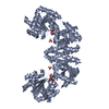
- PDB-3c48: Structure of the retaining glycosyltransferase MshA: The first st... -
+ Open data

- Basic information
| Entry | Database: PDB / ID: 3c48 | ||||||
|---|---|---|---|---|---|---|---|
| Title | Structure of the retaining glycosyltransferase MshA: The first step in mycothiol biosynthesis. Organism: Corynebacterium glutamicum- APO (OPEN) structure. | ||||||
 Components Components | Predicted glycosyltransferases | ||||||
 Keywords Keywords | TRANSFERASE / retaining glycosyltransferase / beta alpha beta / substrate assisted catalysis | ||||||
| Function / homology |  Function and homology information Function and homology informationD-inositol-3-phosphate glycosyltransferase / D-inositol-3-phosphate glycosyltransferase activity / mycothiol biosynthetic process / acetylglucosaminyltransferase activity / magnesium ion binding Similarity search - Function | ||||||
| Biological species |  Corynebacterium glutamicum (bacteria) Corynebacterium glutamicum (bacteria) | ||||||
| Method |  X-RAY DIFFRACTION / SAD / Resolution: 2.1 Å X-RAY DIFFRACTION / SAD / Resolution: 2.1 Å | ||||||
 Authors Authors | Vetting, M.W. / Frantom, P.A. / Blanchard, J.S. | ||||||
 Citation Citation | Journal: J.Biol.Chem. / Year: 2008 Title: Structural and Enzymatic Analysis of MshA from Corynebacterium glutamicum: SUBSTRATE-ASSISTED CATALYSIS Authors: Vetting, M.W. / Frantom, P.A. / Blanchard, J.S. | ||||||
| History |
|
- Structure visualization
| Structure viewer | Molecule: MolmilJmol/JSmol |
|---|
- Downloads & links
-Download
| PDBx/mmCIF format | 3c48.cif.gz | 172.1 KB | Display | PDBx/mmCIF format |
|---|---|---|---|---|
| PDB format | pdb3c48.ent.gz | 135.8 KB | Display | PDB format |
| PDBx/mmJSON format | 3c48.json.gz | Tree view | PDBx/mmJSON format | |
| Others |  Other downloads Other downloads |
-Validation report
| Arichive directory | https://data.pdbj.org/pub/pdb/validation_reports/c4/3c48ftp://data.pdbj.org/pub/pdb/validation_reports/c4/3c48 | HTTPS FTP |
|---|
-Related structure data


-Links
 PDBj
PDBj
- Assembly
| Deposited unit | 
| ||||||||
|---|---|---|---|---|---|---|---|---|---|
| 1 |
| ||||||||
| Unit cell |
|
-Components
| #1: Protein | Mass: 47892.586 Da / Num. of mol.: 2 Source method: isolated from a genetically manipulated source Source: (gene. exp.) Corynebacterium glutamicum (bacteria) / Gene: MshA / Plasmid: pET28 / Production host: References: UniProt: Q8NTA6, Transferases; Glycosyltransferases; Hexosyltransferases #2: Chemical | ChemComp-SO4 /   Mass: 96.063 Da / Num. of mol.: 8 / Source method: obtained synthetically / Formula: SO4 Mass: 96.063 Da / Num. of mol.: 8 / Source method: obtained synthetically / Formula: SO4#3: Water | ChemComp-HOH / |  Mass: 18.015 Da / Num. of mol.: 405 / Source method: isolated from a natural source / Formula: H2O Mass: 18.015 Da / Num. of mol.: 405 / Source method: isolated from a natural source / Formula: H2O |
|---|
-Experimental details
-Experiment
| Experiment | Method: X-RAY DIFFRACTION / Number of used crystals: 1 |
|---|
- Sample preparation
| Crystal | Density Matthews: 2.84 Å3/Da / Density % sol: 56.74 % |
|---|---|
| Crystal grow | Temperature: 291 K / Method: vapor diffusion / pH: 8.5 Details: Protein (15 mg/ml, 400 mM Ammonium sulfate, 10% glycerol, 0.5 mM EDTA, 1 mM BME): Precipitant (20% Peg4000, 100 mM Tris pH 8.5, 200 mm LiSO4), Vapor diffusion under oil, temperature 291K, VAPOR DIFFUSION |
-Data collection
| Diffraction | Mean temperature: 93 K |
|---|---|
| Diffraction source | Source: ROTATING ANODE / Type: RIGAKU RUH3R / Wavelength: 1.5418 Å |
| Detector | Type: RIGAKU RAXIS IV++ / Detector: IMAGE PLATE / Date: Aug 1, 2007 |
| Radiation | Monochromator: MSC Confocal Blue Optics / Protocol: SINGLE WAVELENGTH / Monochromatic (M) / Laue (L): M / Scattering type: x-ray |
| Radiation wavelength | Wavelength: 1.5418 Å / Relative weight: 1 |
| Reflection | Resolution: 2.1→35 Å / Num. all: 178087 / Num. obs: 60789 / % possible obs: 98.7 % / Observed criterion σ(F): 0 / Observed criterion σ(I): 0 / Redundancy: 2.9 % / Biso Wilson estimate: 35.7 Å2 / Rmerge(I) obs: 0.052 / Rsym value: 0.052 / Net I/σ(I): 16.5 |
| Reflection shell | Resolution: 2.1→2.21 Å / Redundancy: 2.5 % / Rmerge(I) obs: 0.238 / Mean I/σ(I) obs: 2.9 / Num. unique all: 8239 / Rsym value: 0.238 / % possible all: 91.5 |
- Processing
| Software |
| |||||||||||||||||||||||||||||||||||||||||||||||||||||||||||||||||||||||||||||||||||||||||||||||||||||||||||||||||||||||||||||
|---|---|---|---|---|---|---|---|---|---|---|---|---|---|---|---|---|---|---|---|---|---|---|---|---|---|---|---|---|---|---|---|---|---|---|---|---|---|---|---|---|---|---|---|---|---|---|---|---|---|---|---|---|---|---|---|---|---|---|---|---|---|---|---|---|---|---|---|---|---|---|---|---|---|---|---|---|---|---|---|---|---|---|---|---|---|---|---|---|---|---|---|---|---|---|---|---|---|---|---|---|---|---|---|---|---|---|---|---|---|---|---|---|---|---|---|---|---|---|---|---|---|---|---|---|---|---|
| Refinement | Method to determine structure: SAD / Resolution: 2.1→34.53 Å / Cor.coef. Fo:Fc: 0.956 / Cor.coef. Fo:Fc free: 0.935 / SU B: 7.904 / SU ML: 0.114 / TLS residual ADP flag: LIKELY RESIDUAL / Cross valid method: THROUGHOUT / σ(F): 0 / σ(I): 0 / ESU R: 0.177 / ESU R Free: 0.161 / Stereochemistry target values: MAXIMUM LIKELIHOOD / Details: HYDROGENS HAVE BEEN ADDED IN THE RIDING POSITIONS
| |||||||||||||||||||||||||||||||||||||||||||||||||||||||||||||||||||||||||||||||||||||||||||||||||||||||||||||||||||||||||||||
| Solvent computation | Ion probe radii: 0.8 Å / Shrinkage radii: 0.8 Å / VDW probe radii: 1.2 Å / Solvent model: MASK | |||||||||||||||||||||||||||||||||||||||||||||||||||||||||||||||||||||||||||||||||||||||||||||||||||||||||||||||||||||||||||||
| Displacement parameters | Biso mean: 36.496 Å2
| |||||||||||||||||||||||||||||||||||||||||||||||||||||||||||||||||||||||||||||||||||||||||||||||||||||||||||||||||||||||||||||
| Refinement step | Cycle: LAST / Resolution: 2.1→34.53 Å
| |||||||||||||||||||||||||||||||||||||||||||||||||||||||||||||||||||||||||||||||||||||||||||||||||||||||||||||||||||||||||||||
| Refine LS restraints |
| |||||||||||||||||||||||||||||||||||||||||||||||||||||||||||||||||||||||||||||||||||||||||||||||||||||||||||||||||||||||||||||
| LS refinement shell | Resolution: 2.1→2.155 Å / Total num. of bins used: 20
| |||||||||||||||||||||||||||||||||||||||||||||||||||||||||||||||||||||||||||||||||||||||||||||||||||||||||||||||||||||||||||||
| Refinement TLS params. | Method: refined / Refine-ID: X-RAY DIFFRACTION
| |||||||||||||||||||||||||||||||||||||||||||||||||||||||||||||||||||||||||||||||||||||||||||||||||||||||||||||||||||||||||||||
| Refinement TLS group |
|
