Mass: 18.015 Da / Num. of mol.: 538 / Source method: isolated from a natural source / Formula: H2O
Has protein modification
Y
Sequence details
THE CONSTRUCT WAS EXPRESSED WITH A PURIFICATION TAG MGSDKIHHHHHHENLYFQG. THE TAG WAS REMOVED WITH ...THE CONSTRUCT WAS EXPRESSED WITH A PURIFICATION TAG MGSDKIHHHHHHENLYFQG. THE TAG WAS REMOVED WITH TEV PROTEASE LEAVING ONLY A GLYCINE (0) FOLLOWED BY THE TARGET SEQUENCE.
-
Experimental details
-
Experiment
Experiment
Method: X-RAY DIFFRACTION / Number of used crystals: 1
-
Sample preparation
Crystal
Density Matthews: 2.84 Å3/Da / Density % sol: 56.69 %
Crystal grow
Temperature: 277 K / Method: vapor diffusion, sitting drop / pH: 8.17 Details: NANODROP, 10.0% PEG 6000, 0.9M Lithium chloride, 0.1M Tris-HCl pH 8.17, VAPOR DIFFUSION, SITTING DROP, temperature 277K
Type: MARMOSAIC 325 mm CCD / Detector: CCD / Date: Dec 5, 2007 / Details: Flat mirror (vertical focusing)
Radiation
Monochromator: Single crystal Si(111) bent (horizontal focusing) Protocol: MAD / Monochromatic (M) / Laue (L): M / Scattering type: x-ray
Radiation wavelength
ID
Wavelength (Å)
Relative weight
1
0.91837
1
2
0.97951
1
Reflection
Resolution: 1.9→29.709 Å / Num. obs: 95241 / % possible obs: 99.9 % / Observed criterion σ(I): -3 / Biso Wilson estimate: 27.097 Å2 / Rmerge(I) obs: 0.068 / Net I/σ(I): 9.14
Reflection shell
Resolution (Å)
Rmerge(I) obs
Mean I/σ(I) obs
Num. measured obs
Num. unique obs
Diffraction-ID
% possible all
1.9-1.97
0.534
2.2
57286
18305
1
99.7
1.97-2.05
0.451
2.6
57488
18056
1
100
2.05-2.14
0.378
3.1
55023
17340
1
100
2.14-2.25
0.292
4
55527
17410
1
100
2.25-2.39
0.224
5.2
57441
17944
1
100
2.39-2.58
0.156
7.2
59311
18464
1
100
2.58-2.84
0.106
10
57412
17851
1
100
2.84-3.25
0.071
14
56955
17849
1
99.9
3.25-4.08
0.046
19.9
55287
17720
1
99.9
4.08-29.709
0.034
23.1
55359
18040
1
99.1
-
Phasing
Phasing
Method: MAD
-
Processing
Software
Name
Version
Classification
NB
REFMAC
5.4.0067
refinement
PHENIX
refinement
SOLVE
phasing
MolProbity
3beta29
modelbuilding
XSCALE
datascaling
PDB_EXTRACT
3
dataextraction
MAR345
CCD
datacollection
XDS
datareduction
Refinement
Method to determine structure: MAD / Resolution: 1.9→29.709 Å / Cor.coef. Fo:Fc: 0.969 / Cor.coef. Fo:Fc free: 0.949 / SU B: 7.92 / SU ML: 0.112 / TLS residual ADP flag: LIKELY RESIDUAL / Cross valid method: THROUGHOUT / σ(F): 0 / ESU R: 0.129 / ESU R Free: 0.132 Stereochemistry target values: MAXIMUM LIKELIHOOD WITH PHASES Details: 1. HYDROGENS HAVE BEEN ADDED IN THE RIDING POSITIONS. 2. A MET-INHIBITION PROTOCOL WAS USED FOR SELENOMETHIONINE INCORPORATION DURING PROTEIN EXPRESSION. THE OCCUPANCY OF THE SE ATOMS IN THE ...Details: 1. HYDROGENS HAVE BEEN ADDED IN THE RIDING POSITIONS. 2. A MET-INHIBITION PROTOCOL WAS USED FOR SELENOMETHIONINE INCORPORATION DURING PROTEIN EXPRESSION. THE OCCUPANCY OF THE SE ATOMS IN THE MSE RESIDUES WAS REDUCED TO 0.75 FOR THE REDUCED SCATTERING POWER DUE TO PARTIAL S-MET INCORPORATION. 3. ATOM RECORD CONTAINS RESIDUAL B FACTORS ONLY. 4. CL AND GOL WERE MODELED BASED ON CRYSTALLIZATION AND CRYOPROTECTION CONDITIONS.
Rfactor
Num. reflection
% reflection
Selection details
Rfree
0.233
4765
5 %
RANDOM
Rwork
0.183
-
-
-
obs
0.186
95092
99.93 %
-
Solvent computation
Ion probe radii: 0.8 Å / Shrinkage radii: 0.8 Å / VDW probe radii: 1.2 Å / Solvent model: MASK
Displacement parameters
Biso mean: 29.67 Å2
Baniso -1
Baniso -2
Baniso -3
1-
0.66 Å2
0.33 Å2
0 Å2
2-
-
0.66 Å2
0 Å2
3-
-
-
-0.99 Å2
Refinement step
Cycle: LAST / Resolution: 1.9→29.709 Å
Protein
Nucleic acid
Ligand
Solvent
Total
Num. atoms
7141
0
27
538
7706
Refine LS restraints
Refine-ID
Type
Dev ideal
Dev ideal target
Number
X-RAY DIFFRACTION
r_bond_refined_d
0.014
0.021
7447
X-RAY DIFFRACTION
r_bond_other_d
0.001
0.02
5091
X-RAY DIFFRACTION
r_angle_refined_deg
1.294
1.957
10131
X-RAY DIFFRACTION
r_angle_other_deg
1.418
3
12338
X-RAY DIFFRACTION
r_dihedral_angle_1_deg
4.453
5
912
X-RAY DIFFRACTION
r_dihedral_angle_2_deg
34.971
22.834
374
X-RAY DIFFRACTION
r_dihedral_angle_3_deg
13.002
15
1322
X-RAY DIFFRACTION
r_dihedral_angle_4_deg
14.234
15
73
X-RAY DIFFRACTION
r_chiral_restr
0.077
0.2
1145
X-RAY DIFFRACTION
r_gen_planes_refined
0.007
0.02
8233
X-RAY DIFFRACTION
r_gen_planes_other
0.002
0.02
1605
X-RAY DIFFRACTION
r_mcbond_it
1.978
3
4398
X-RAY DIFFRACTION
r_mcbond_other
0.553
3
1773
X-RAY DIFFRACTION
r_mcangle_it
3.351
5
7153
X-RAY DIFFRACTION
r_scbond_it
5.593
8
3049
X-RAY DIFFRACTION
r_scangle_it
8.326
11
2955
LS refinement shell
Resolution: 1.9→1.949 Å / Total num. of bins used: 20
Rfactor
Num. reflection
% reflection
Rfree
0.371
350
-
Rwork
0.317
6499
-
all
-
6849
-
obs
-
-
99.93 %
Refinement TLS params.
Method: refined / Refine-ID: X-RAY DIFFRACTION
ID
L11 (°2)
L12 (°2)
L13 (°2)
L22 (°2)
L23 (°2)
L33 (°2)
S11 (Å °)
S12 (Å °)
S13 (Å °)
S21 (Å °)
S22 (Å °)
S23 (Å °)
S31 (Å °)
S32 (Å °)
S33 (Å °)
T11 (Å2)
T12 (Å2)
T13 (Å2)
T22 (Å2)
T23 (Å2)
T33 (Å2)
Origin x (Å)
Origin y (Å)
Origin z (Å)
1
0.422
-0.1185
-0.2021
0.1241
-0.0685
0.5972
-0.0534
0.0677
-0.019
0.0047
-0.0249
0.0196
0.0734
-0.1451
0.0783
-0.0266
0.005
0.0457
-0.0205
-0.0068
-0.0107
11.7375
54.1706
31.4677
2
0.2518
-0.1783
0.026
0.3785
-0.1472
0.597
-0.0276
0.1091
0.0566
-0.0708
-0.0577
-0.0773
-0.1984
0.0118
0.0853
0.0058
0.0249
0.0187
-0.0145
0.0363
-0.0587
22.4269
68.7886
24.3031
3
0.1138
-0.0105
0.1996
0.0279
0.1283
1.1469
0.0247
0.0137
-0.0153
0.0246
-0.0492
0.0352
0.3304
0.0054
0.0246
0.0925
0.0423
0.044
-0.1114
0.0055
0.0277
5.7658
86.8911
73.4796
Refinement TLS group
ID
Refine-ID
Refine TLS-ID
Auth asym-ID
Label asym-ID
Auth seq-ID
Label seq-ID
1
X-RAY DIFFRACTION
1
A
A
0 - 289
1 - 290
2
X-RAY DIFFRACTION
2
B
B
0 - 288
1 - 289
3
X-RAY DIFFRACTION
3
C
C
0 - 288
1 - 289
+
About Yorodumi
-
News
-
Feb 9, 2022. New format data for meta-information of EMDB entries
New format data for meta-information of EMDB entries
Version 3 of the EMDB header file is now the official format.
The previous official version 1.9 will be removed from the archive.
In the structure databanks used in Yorodumi, some data are registered as the other names, "COVID-19 virus" and "2019-nCoV". Here are the details of the virus and the list of structure data.
Jan 31, 2019. EMDB accession codes are about to change! (news from PDBe EMDB page)
EMDB accession codes are about to change! (news from PDBe EMDB page)
The allocation of 4 digits for EMDB accession codes will soon come to an end. Whilst these codes will remain in use, new EMDB accession codes will include an additional digit and will expand incrementally as the available range of codes is exhausted. The current 4-digit format prefixed with “EMD-” (i.e. EMD-XXXX) will advance to a 5-digit format (i.e. EMD-XXXXX), and so on. It is currently estimated that the 4-digit codes will be depleted around Spring 2019, at which point the 5-digit format will come into force.
The EM Navigator/Yorodumi systems omit the EMD- prefix.
Related info.:Q: What is EMD? / ID/Accession-code notation in Yorodumi/EM Navigator
Yorodumi is a browser for structure data from EMDB, PDB, SASBDB, etc.
This page is also the successor to EM Navigator detail page, and also detail information page/front-end page for Omokage search.
The word "yorodu" (or yorozu) is an old Japanese word meaning "ten thousand". "mi" (miru) is to see.
Related info.:EMDB / PDB / SASBDB / Comparison of 3 databanks / Yorodumi Search / Aug 31, 2016. New EM Navigator & Yorodumi / Yorodumi Papers / Jmol/JSmol / Function and homology information / Changes in new EM Navigator and Yorodumi
 Movie
Movie Controller

 Yorodumi
 Open data
 Basic information
 Components
 Keywords
 Function and homology information
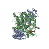
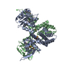
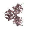
 Exiguobacterium sibiricum (bacteria)
 X-RAY DIFFRACTION /
  Authors
 Citation
 Structure visualization
 Downloads & links
 Other downloads

 PDBj
PDBj Assembly
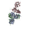

 Mass: 92.094 Da / Num. of mol.: 4 / Source method: obtained synthetically / Formula: C3H8O3
 Mass: 35.453 Da / Num. of mol.: 3 / Source method: obtained synthetically / Formula: Cl
 Mass: 18.015 Da / Num. of mol.: 538 / Source method: isolated from a natural source / Formula: H2O
 Sample preparation
 / Beamline: BL11-1 / Wavelength: 0.91837, 0.97951
 Processing