 Movie
Movie Controller

[English] 日本語
 Yorodumi
- PDB-3bz6: Crystal structure of a conserved protein of unknown function from... -
+ Open data

- Basic information
| Entry | Database: PDB / ID: 3bz6 | |||||||||
|---|---|---|---|---|---|---|---|---|---|---|
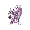
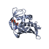
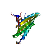
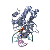
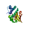
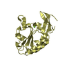
| Title | Crystal structure of a conserved protein of unknown function from Pseudomonas syringae pv. tomato str. DC3000 | |||||||||
 Components Components | UPF0502 protein PSPTO_2686 | |||||||||
 Keywords Keywords | STRUCTURAL GENOMICS / UNKNOWN FUNCTION / APC84902 / conserved domain / Pseudomonas syringae pv. tomato str. DC3000 / PSI-2 / Protein Structure Initiative / Midwest Center for Structural Genomics / MCSG | |||||||||
| Function / homology | Protein of unknown function DUF480 / Protein of unknown function, DUF480 / Winged helix-like DNA-binding domain superfamily/Winged helix DNA-binding domain / Arc Repressor Mutant, subunit A / Winged helix DNA-binding domain superfamily / Winged helix-like DNA-binding domain superfamily / Orthogonal Bundle / Mainly Alpha / UPF0502 protein PSPTO_2686 Function and homology information Function and homology information | |||||||||
| Biological species |  Pseudomonas syringae pv. tomato (bacteria) Pseudomonas syringae pv. tomato (bacteria) | |||||||||
| Method |  X-RAY DIFFRACTION / SYNCHROTRON / MAD / Resolution: 2.21 Å X-RAY DIFFRACTION / SYNCHROTRON / MAD / Resolution: 2.21 Å | |||||||||
 Authors Authors | Tan, K. / Duggan, E. / Clancy, S. / Joachimiak, A. / Midwest Center for Structural Genomics (MCSG) | |||||||||
 Citation Citation | Journal: To be Published Title: The crystal structure of a conserved protein of unknown function from Pseudomonas syringae pv. tomato str. DC3000. Authors: Tan, K. / Duggan, E. / Clancy, S. / Joachimiak, A. | |||||||||
| History |
|
- Structure visualization
| Structure viewer | Molecule: MolmilJmol/JSmol |
|---|
- Downloads & links
-Download
| PDBx/mmCIF format | 3bz6.cif.gz | 46.7 KB | Display | PDBx/mmCIF format |
|---|---|---|---|---|
| PDB format | pdb3bz6.ent.gz | 32.9 KB | Display | PDB format |
| PDBx/mmJSON format | 3bz6.json.gz | Tree view | PDBx/mmJSON format | |
| Others |  Other downloads Other downloads |
-Validation report
| Arichive directory | https://data.pdbj.org/pub/pdb/validation_reports/bz/3bz6ftp://data.pdbj.org/pub/pdb/validation_reports/bz/3bz6 | HTTPS FTP |
|---|
-Related structure data
| Similar structure data | |
|---|---|
| Other databases |


-Links
 PDBj
PDBj- Assembly
| Deposited unit | 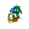
| ||||||||
|---|---|---|---|---|---|---|---|---|---|
| 1 |
| ||||||||
| Unit cell |
| ||||||||
| Details | AUTHORS STATE THAT THE BIOLOGICAL UNIT OF THIS PROTEIN IS EXPERIMENTALLY UNKNOWN. IT IS LIKELY A MONOMER. |
-Components
| #1: Protein | Mass: 20532.506 Da / Num. of mol.: 1 / Fragment: Residues 1-180 Source method: isolated from a genetically manipulated source Source: (gene. exp.) Pseudomonas syringae pv. tomato (bacteria)Species: Pseudomonas syringae group genomosp. 3 / Strain: DC3000 / Gene: PSPTO_2686 / Plasmid: pMCSG7 / Species (production host): Escherichia coli / Production host: |
|---|---|
| #2: Water | ChemComp-HOH /  Mass: 18.015 Da / Num. of mol.: 26 / Source method: isolated from a natural source / Formula: H2O Mass: 18.015 Da / Num. of mol.: 26 / Source method: isolated from a natural source / Formula: H2O |
| Has protein modification | Y |
-Experimental details
-Experiment
| Experiment | Method: X-RAY DIFFRACTION / Number of used crystals: 1 |
|---|
- Sample preparation
| Crystal | Density Matthews: 2.67 Å3/Da / Density % sol: 53.87 % |
|---|---|
| Crystal grow | Temperature: 277 K / Method: vapor diffusion, sitting drop Details: 0.1M Sodium acetate, 20% Isopropanol, 20% PEG 4000, VAPOR DIFFUSION, SITTING DROP, temperature 277K |
-Data collection
| Diffraction | Mean temperature: 100 K | |||||||||
|---|---|---|---|---|---|---|---|---|---|---|
| Diffraction source | Source: SYNCHROTRON / Site: APS  / Beamline: 19-ID / Wavelength: 0.97929, 0.97943 / Beamline: 19-ID / Wavelength: 0.97929, 0.97943 | |||||||||
| Detector | Type: ADSC QUANTUM 315 / Detector: CCD / Date: Aug 13, 2006 / Details: Mirrors | |||||||||
| Radiation | Monochromator: Si 111 Crystal / Protocol: MAD / Monochromatic (M) / Laue (L): M / Scattering type: x-ray | |||||||||
| Radiation wavelength |
| |||||||||
| Reflection | Resolution: 2.2→31.54 Å / Num. all: 10229 / Num. obs: 10229 / % possible obs: 93.1 % / Observed criterion σ(F): 0 / Observed criterion σ(I): 0 / Redundancy: 13.2 % / Rmerge(I) obs: 0.091 / Net I/σ(I): 46 | |||||||||
| Reflection shell | Resolution: 2.2→2.25 Å / Redundancy: 9.5 % / Rmerge(I) obs: 0.391 / Mean I/σ(I) obs: 2.59 / Num. unique all: 426 / % possible all: 58.5 |
- Processing
| Software |
| ||||||||||||||||||||||||||||||||||||||||||||||||||||||||||||||||||||||||||||||||||||||||||||||||||||||||||||||||||||||||||||||||||||||||||||||||||||||||||||||||||||||||||
|---|---|---|---|---|---|---|---|---|---|---|---|---|---|---|---|---|---|---|---|---|---|---|---|---|---|---|---|---|---|---|---|---|---|---|---|---|---|---|---|---|---|---|---|---|---|---|---|---|---|---|---|---|---|---|---|---|---|---|---|---|---|---|---|---|---|---|---|---|---|---|---|---|---|---|---|---|---|---|---|---|---|---|---|---|---|---|---|---|---|---|---|---|---|---|---|---|---|---|---|---|---|---|---|---|---|---|---|---|---|---|---|---|---|---|---|---|---|---|---|---|---|---|---|---|---|---|---|---|---|---|---|---|---|---|---|---|---|---|---|---|---|---|---|---|---|---|---|---|---|---|---|---|---|---|---|---|---|---|---|---|---|---|---|---|---|---|---|---|---|---|---|
| Refinement | Method to determine structure: MAD / Resolution: 2.21→31.54 Å / Cor.coef. Fo:Fc: 0.963 / Cor.coef. Fo:Fc free: 0.955 / SU B: 17.474 / SU ML: 0.195 / TLS residual ADP flag: LIKELY RESIDUAL / Cross valid method: THROUGHOUT / σ(F): 0 / σ(I): 0 / ESU R: 0.252 / ESU R Free: 0.207 / Stereochemistry target values: MAXIMUM LIKELIHOOD / Details: HYDROGENS HAVE BEEN ADDED IN THE RIDING POSITIONS
| ||||||||||||||||||||||||||||||||||||||||||||||||||||||||||||||||||||||||||||||||||||||||||||||||||||||||||||||||||||||||||||||||||||||||||||||||||||||||||||||||||||||||||
| Solvent computation | Ion probe radii: 0.8 Å / Shrinkage radii: 0.8 Å / VDW probe radii: 1.2 Å / Solvent model: MASK | ||||||||||||||||||||||||||||||||||||||||||||||||||||||||||||||||||||||||||||||||||||||||||||||||||||||||||||||||||||||||||||||||||||||||||||||||||||||||||||||||||||||||||
| Displacement parameters | Biso mean: 58.166 Å2
| ||||||||||||||||||||||||||||||||||||||||||||||||||||||||||||||||||||||||||||||||||||||||||||||||||||||||||||||||||||||||||||||||||||||||||||||||||||||||||||||||||||||||||
| Refinement step | Cycle: LAST / Resolution: 2.21→31.54 Å
| ||||||||||||||||||||||||||||||||||||||||||||||||||||||||||||||||||||||||||||||||||||||||||||||||||||||||||||||||||||||||||||||||||||||||||||||||||||||||||||||||||||||||||
| Refine LS restraints |
| ||||||||||||||||||||||||||||||||||||||||||||||||||||||||||||||||||||||||||||||||||||||||||||||||||||||||||||||||||||||||||||||||||||||||||||||||||||||||||||||||||||||||||
| LS refinement shell | Resolution: 2.21→2.26 Å / Total num. of bins used: 20
| ||||||||||||||||||||||||||||||||||||||||||||||||||||||||||||||||||||||||||||||||||||||||||||||||||||||||||||||||||||||||||||||||||||||||||||||||||||||||||||||||||||||||||
| Refinement TLS params. | Method: refined / Origin x: -9.385 Å / Origin y: 32.111 Å / Origin z: 6.325 Å
|
