 Movie
Movie Controller

[English] 日本語
 Yorodumi
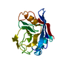
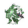
- PDB-3bt8: Crystal Structure of Mutant Cyclophilin (R147A) from Leishmania d... -
+ Open data

- Basic information
| Entry | Database: PDB / ID: 3bt8 | ||||||
|---|---|---|---|---|---|---|---|
| Title | Crystal Structure of Mutant Cyclophilin (R147A) from Leishmania donovani | ||||||
 Components Components | Peptidyl-prolyl cis-trans isomerase | ||||||
 Keywords Keywords | ISOMERASE / CYCLOPHILIN / ROTAMASE / PROLINE / CIS-TRANS / PROTOZOA / LEISHMANIA / DONOVANI / KALA-AZAR | ||||||
| Function / homology |  Function and homology information Function and homology informationcyclosporin A binding / peptidylprolyl isomerase / peptidyl-prolyl cis-trans isomerase activity / protein folding / intracellular membrane-bounded organelle / cytoplasm Similarity search - Function | ||||||
| Biological species |  Leishmania donovani (eukaryote) Leishmania donovani (eukaryote) | ||||||
| Method |  X-RAY DIFFRACTION / MOLECULAR REPLACEMENT / Resolution: 2.7 Å X-RAY DIFFRACTION / MOLECULAR REPLACEMENT / Resolution: 2.7 Å | ||||||
 Authors Authors | Venugopal, V. / Sen, B. / Datta, A.K. / Banerjee, R. | ||||||
 Citation Citation | Journal: To be Published Title: Crystal Structure of Mutant Cyclophilin from Leishmania Donovani Authors: Venugopal, V. / Sen, B. / Datta, A.K. / Banerjee, R. #1: Journal: Acta Crystallogr.,Sect.F / Year: 2007Title: Structure of cyclophilin from Leishmania donovani at 1.97 A resolution Authors: Venugopal, V. / Sen, B. / Datta, A.K. / Banerjee, R. #2: Journal: Biochemistry / Year: 2007 Title: Amino acid residues of Leishmania donovani cyclophilin key to interaction with its adenosine kinase: biological implications Authors: Sen, B. / Venugopal, V. / Chakraborty, A. / Datta, R. / Dolai, S. / Banerjee, R. / Datta, A.K. | ||||||
| History |
|
- Structure visualization
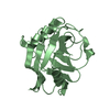
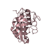
| Structure viewer | Molecule: MolmilJmol/JSmol |
|---|
- Downloads & links
-Download
| PDBx/mmCIF format | 3bt8.cif.gz | 46 KB | Display | PDBx/mmCIF format |
|---|---|---|---|---|
| PDB format | pdb3bt8.ent.gz | 31.4 KB | Display | PDB format |
| PDBx/mmJSON format | 3bt8.json.gz | Tree view | PDBx/mmJSON format | |
| Others |  Other downloads Other downloads |
-Validation report
| Summary document | 3bt8_validation.pdf.gz | 421.6 KB | Display | wwPDB validaton report |
|---|---|---|---|---|
| Full document | 3bt8_full_validation.pdf.gz | 424.9 KB | Display | |
| Data in XML | 3bt8_validation.xml.gz | 8.8 KB | Display | |
| Data in CIF | 3bt8_validation.cif.gz | 11.1 KB | Display | |
| Arichive directory | https://data.pdbj.org/pub/pdb/validation_reports/bt/3bt8ftp://data.pdbj.org/pub/pdb/validation_reports/bt/3bt8 | HTTPS FTP |
-Related structure data
| Related structure data |  2haqS S: Starting model for refinement |
|---|---|
| Similar structure data |


-Links
 PDBj
PDBj

- Assembly
| Deposited unit | 
| ||||||||
|---|---|---|---|---|---|---|---|---|---|
| 1 |
| ||||||||
| Unit cell |
|
-Components
| #1: Protein | Mass: 19004.426 Da / Num. of mol.: 1 / Fragment: UNP residues 22-187 / Mutation: R147A Source method: isolated from a genetically manipulated source Source: (gene. exp.) Leishmania donovani (eukaryote) / Plasmid: pQE32 / Production host:  |
|---|---|
| #2: Water | ChemComp-HOH /  Mass: 18.015 Da / Num. of mol.: 38 / Source method: isolated from a natural source / Formula: H2O Mass: 18.015 Da / Num. of mol.: 38 / Source method: isolated from a natural source / Formula: H2O |
-Experimental details
-Experiment
| Experiment | Method: X-RAY DIFFRACTION / Number of used crystals: 1 |
|---|
- Sample preparation
| Crystal | Density Matthews: 2.62 Å3/Da / Density % sol: 53.02 % |
|---|---|
| Crystal grow | Temperature: 292 K / Method: vapor diffusion, hanging drop / pH: 8.5 Details: 0.01M TRIS, 10.0% PEG 3350, 0.02% AZIDE, PH 8.5, VAPOR DIFFUSION, HANGING DROP, temperature 292K |
-Data collection
| Diffraction | Mean temperature: 293 K |
|---|---|
| Diffraction source | Source: ROTATING ANODE / Type: RIGAKU RU200 / Wavelength: 1.5418 Å |
| Detector | Type: MARRESEARCH / Detector: IMAGE PLATE / Date: Sep 1, 2007 / Details: OSMIC MAXFLUX CONFOCAL MULTILAYER MIRRORS |
| Radiation | Protocol: SINGLE WAVELENGTH / Monochromatic (M) / Laue (L): M / Scattering type: x-ray |
| Radiation wavelength | Wavelength: 1.5418 Å / Relative weight: 1 |
| Reflection | Resolution: 2.7→20 Å / Num. obs: 5450 / % possible obs: 99.5 % / Redundancy: 6.15 % / Biso Wilson estimate: 4.7 Å2 / Rmerge(I) obs: 0.05 / Net I/σ(I): 11 |
| Reflection shell | Resolution: 2.7→2.75 Å / Redundancy: 6.17 % / Rmerge(I) obs: 0.152 / Mean I/σ(I) obs: 4.2 / % possible all: 99.3 |
- Processing
| Software |
| ||||||||||||||||||||
|---|---|---|---|---|---|---|---|---|---|---|---|---|---|---|---|---|---|---|---|---|---|
| Refinement | Method to determine structure: MOLECULAR REPLACEMENT Starting model: PDB ENTRY 2HAQ Resolution: 2.7→17.3 Å / Rfactor Rfree error: 0.012 / Isotropic thermal model: RESTRAINED / Cross valid method: THROUGHOUT / σ(F): 2
| ||||||||||||||||||||
| Displacement parameters | Biso mean: 29.3 Å2
| ||||||||||||||||||||
| Refine analyze |
| ||||||||||||||||||||
| Refinement step | Cycle: LAST / Resolution: 2.7→17.3 Å
| ||||||||||||||||||||
| Refine LS restraints |
| ||||||||||||||||||||
| LS refinement shell | Resolution: 2.7→2.87 Å / Rfactor Rfree error: 0.031 / Total num. of bins used: 6
| ||||||||||||||||||||
| Xplor file |
|
